乳腺癌是最常见的恶性肿瘤之一,全世界每年新增约100万患者,其中以女性为主,占女性恶性肿瘤总数的18%[1-2]。随着超声技术的发展, 及超声分辨率不断提高,乳腺肿瘤的显示率也越来越高,超声可以观察乳腺肿块的细微形态特征,了解侵袭转移、血管生成等生物学行为。自动乳腺全容积成像(automated breast volume scanner,ABVS)是一项新型的超声检查技术,与常规二维超声比较,其优势在于能展示乳腺的冠状切面,可为临床提供更多的诊断及治疗信息[3-8]。ABVS冠状切面汇聚征是乳腺癌常见的典型影像特征,而肿瘤的分子基础决定生物学行为和影像学表现。至今,国内外关于乳腺浸润性导管癌(invasive ductal breast carcinoma,IDBC)患者ABVS冠状面的汇聚征与肿瘤临床病理变量的相关性研究较少,本研究旨在通过分析IDBC患者的ABVS冠状切面汇聚征这一特征与肿瘤临床病理变量之间的关系,为乳腺癌的预后评估和选择临床治疗方案提供影像学依据。
1 资料与方法
1.1 一般资料
收集2015年2月—2017年1月在中南大学湘雅三医院行手术治疗的IDBC的女性患者160例,共计160个肿块作为研究对象。有多个肿块的患者仅取较大肿块作为分析对象。纳入标准:⑴ 患者术前均行常规二维超声及ABVS检查且经术后病理证实为IDBC;⑵ 术前未进行过放、化疗等治疗的原发性IDBC患者。排除标准:⑴ 术后病理显示合并其他肿瘤组织类型者;⑵ 同时患有其他器官、组织恶性疾病者。160例IDBC患者的年龄范围为28~76岁,平均(49.5±5.4)岁。
1.2 常规二维超声扫查
使用SIEMENS ACUSON S2000彩色多普勒超声检查仪,频率范围5.0~14 MHz,扫查探头9L4。嘱受检者平卧位,保持双臂外展以使双乳和腋窝充分暴露。以乳头为中心分别进行纵切、横切和斜切高频二维超声有序扫查,并扫查双侧腋下淋巴结。一旦探查到肿块,则记录肿块的直径、内部回声、边缘、有无包膜、内部有无钙化、纵横比、后方有无衰减及病变与周围组织关系的情况等声像图特性。并使用彩色多普勒血流成像(color doppler flowing imaging,CDFI)对肿块内部和其周围血运情况进行观察。
1.3 ABVS扫查
SIEMENS ACUSON S2000 ABVS采集系统,包括可调节的机械臂、触屏显示器和扫描盒,其扫描探头为高频线阵探头,频率5~14 MHz。采集到的图像层厚常设定为0.5 mm。行ABVS扫查前,嘱患者保持平卧位,根据受检者乳房的大小选择设备的最优扫查预设条件,再调节机械臂,施加恰当的压力,让探头紧紧贴于乳腺皮肤层,使声像图显示最为清晰。扫描探头对每一乳腺以内侧位、外侧位及正位有序扫查,而乳头均包含在每次扫查平面内,扫查每一方位时间约需55~65 s。每一次扫查结束后,在显示屏上标记乳头并保存,数据自动传输至影像数据处理系统进行三维重建,获取A B V S声像图供研究人员对图像反复阅读。本研究中所有图像的采集由1名有2年ABVS工作经验的初级医生完成,再由1名初级医师及1名副主任医师(工作时间分别为3年、20年)共同阅读A B V S声像图。汇聚征诊断标准参照报道[6],当条索状中等回声或高回声从肿块周围向肿块呈星芒状聚集,出现于全部或部分A B V S冠状面图像上时则称为汇聚征(图1 A-B);无汇聚征者表现为:肿块边界清晰,周边回声均匀,无放射状改变(图1C-D)。由2名医师读片前对患者的相关病史、其他影像学检查结果及病理结果不知情。当2名医师观察结果不一致时,则通过讨论后达成共识。
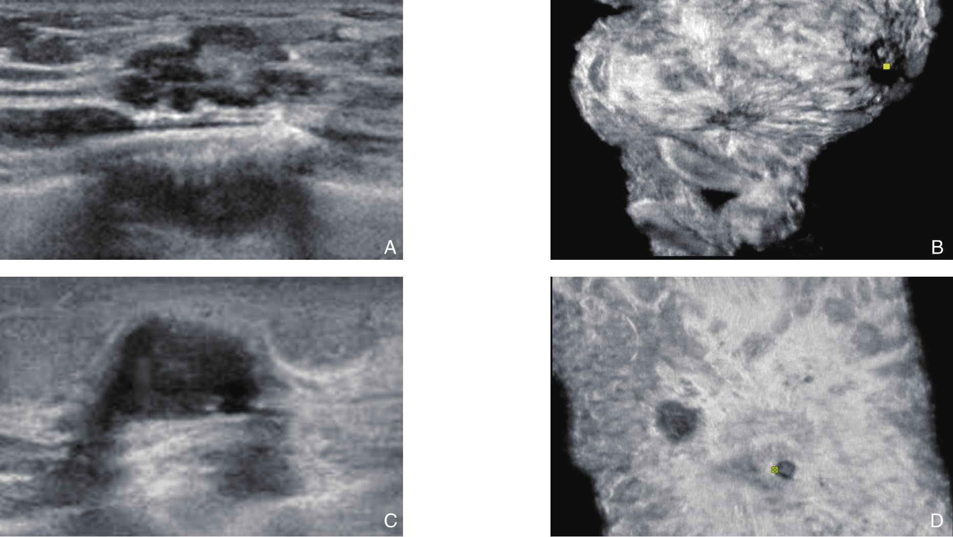
图1 IDBC肿块的常规二维超声及ABVS图像 A:常规二维超声显示乳腺肿块边界不清,呈毛刺征;B:ABVS冠状面可见汇聚征;C:二维超声示乳腺肿块边界清晰;D:ABVS冠状面乳腺肿块未见明显汇聚征
Figure1 Conventional two-dimensional ultrasound and ABVS images of IDBC A: The obscure border of the breast mass and spicule sign showed in conventional two-dimensional ultrasound; B: The convergence sign showed in the coronal plane of ABVS; C: The clear border of the breast mass showed in two-dimensional ultrasound; D: No convergence sign showed in the coronal plane of ABVS
1.4 免疫组化实验及结果判断
免疫组化试剂材料:兔抗人雌激素受体(estrogen receptor,ER)单克隆抗体、鼠抗人孕激素受体(progesterone receptor,PR)单克隆抗体、鼠抗人人表皮生长因子受体(human epidermal growth factor receptor 2,Her-2) 单克隆抗体、鼠抗人Ki-67抗体、鼠抗人表皮生长因子受体(epidermal growth factor receptor,EGFR)抗体。其他包括:胎牛血清1 mol/L的TBS缓冲液0.01 mmol/L柠檬酸盐缓冲液(CB,pH6.0,1000 mL)等。将乳腺肿瘤组织用甲醛固定,石蜡包埋切片,实验步骤严格按照试剂盒规程要求进行染色操作。
免疫组化判定标准[9-12]:ER、PR、Ki-67表达阳性主要体现为细胞核染色,Her-2及EGFR表达阳性染色则主要体现为胞膜或胞质染色。ER或PR:染色细胞数量≥10%为阳性(+),<10%为阴性(-)。Her-2表达分为0,1+,2+或3+,将0或者1+定义为Her-2(-),3+定义为Her-2(+),当表达为2+时应行荧光免疫杂交(fluorescence in situ hybridization,FISH)检测,存在基因拷贝数扩增者则为Her-2(+)。Ki-67:染色细胞数量≥14%为阳性(+),<14%为阴性(-)。EGFR:染色细胞数量>10%为阳性(+),<10%为阴性(-)。
1.5 统计学处理
本研究数据应用SPSS 16.0统计软件分析,计量资料使用均数±标准差( ±s)表示,两组间的计数资料则采用χ2检验。P<0.05为差异有统计学意义。
2 结 果
2.1 IDBC肿块的常规二维超声及ABVS特征
160例IDBC肿块中,形态规则86例,不规则74例;边界清晰94例,不清晰66例;内部回声均匀62例,不均匀98例;内部有微钙化95例,无微钙化65例;后方有衰减有48例,无112例;导管有病变43例,无病变117例;具有汇聚征56例(35.0%),无汇聚征104例(65.0%)。160例IDBC肿块常规二维超声及ABVS特征如下(表1)。
表1 160例IDBC肿块的常规二维超声及ABVS特征[n(%)]
Table1 The imaging features of conventional two-dimensional ultrasound and ABVS in the 160 IDBC masses [n (%)]
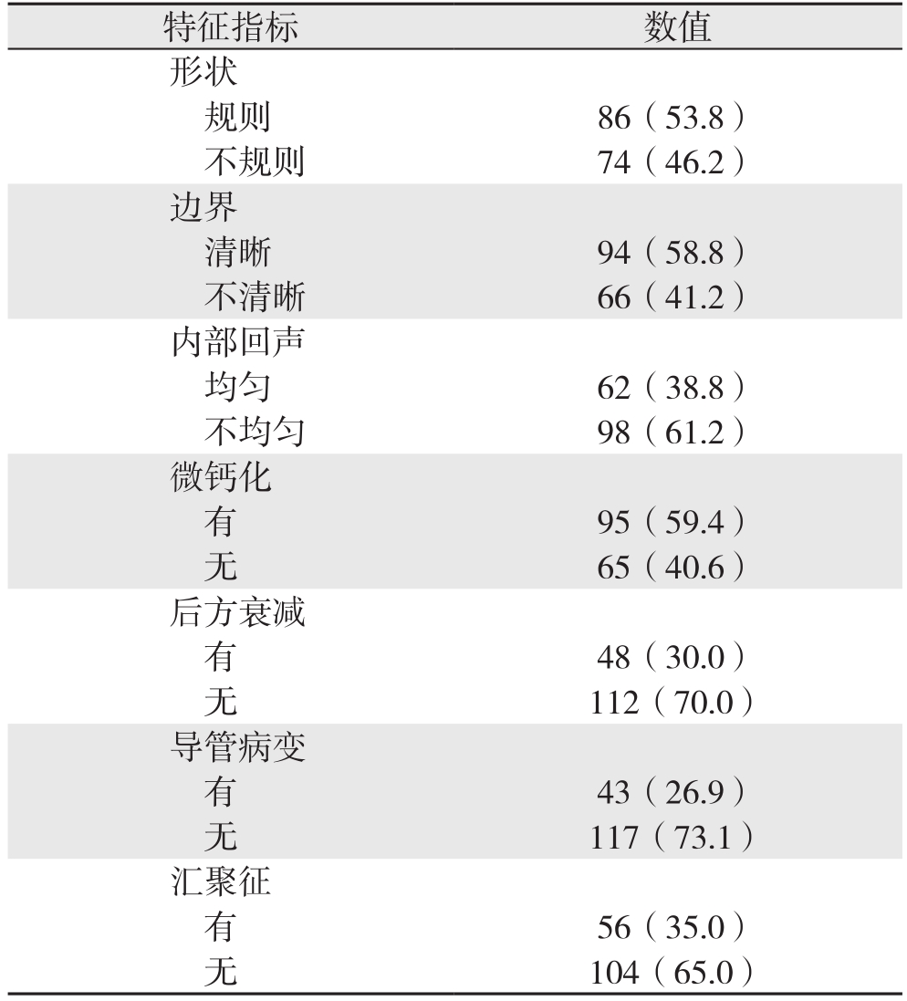
特征指标 数值形状规则 86(53.8)不规则 74(46.2)边界清晰 94(58.8)不清晰 66(41.2)内部回声均匀 62(38.8)不均匀 98(61.2)微钙化有95(59.4)无65(40.6)后方衰减有48(30.0)无112(70.0)导管病变有43(26.9)无117(73.1)汇聚征有56(35.0)无104(65.0)
2.2 IDBC肿块的术后病理结果
160例I D B C肿块中,K i-67(+)78例(48.8%),K i-67(-)82例(51.2%);EGFR(+)61例(38.1%),EGFR(-)99例(61.9%);ER(+)112例(70.0%),ER(-)48例(30.0%);PR(+)98例(61.3%),PR(-)62例(38.7%);Her-2(+)106例(66.3%),Her-2(-)54例(33.7%)(图2-3)。
2.3 汇聚征与IDBC患者的临床特征的关系
依据前述关于ABVS冠状面汇聚征的定义,将160例IDBC肿块分为汇聚征组与无汇聚征组。汇聚征在组织学分级I~II级组和腋窝淋巴结转移组中出现的比例高,差异有统计学意义(均P<0.05)。汇聚征在不同年龄、不同肿块大小、不同绝经状态组中出现比例差异无统计学意义(均P>0.05)(表2)。
2.4 IDBC肿块的汇聚征与肿瘤分子标记物的关系
160例I D B C肿块中,汇聚征在E R阳性、PR阳性、EGFR阴性、Ki-67阴性组中出现的比例高于无汇聚征组,且差异有统计学差异(均P<0.05);汇聚征在不同Her-2状态组中出现的比例差异无统计学差异(P>0.05)(表3)。
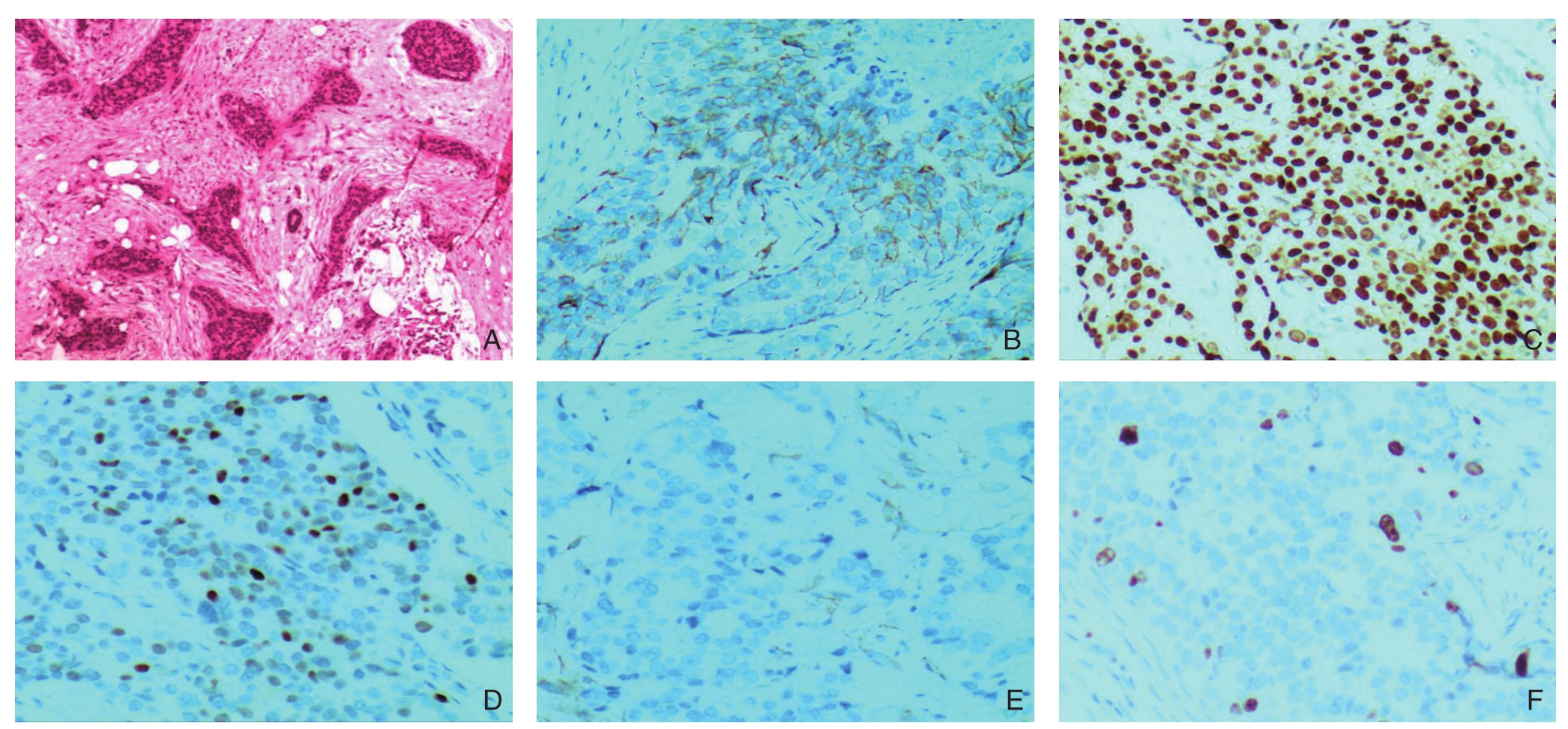
图2 有汇聚征表现的IDBC肿块的术后病理情况(×400) A:病理结果为IDBC(HE染色);B:Her-2(+);C:ER(+);D:PR(+);E:EGFR(-);F:Ki-67(-)
Figure2 The postoperative pathology of IDBC mass showing convergence sign (×400) A: Pathological feature of IDBC (HE staining);B: Her-2 (+); C: ER (+); D: PR (+); E: EGFR (–); F: ki-67 (–)
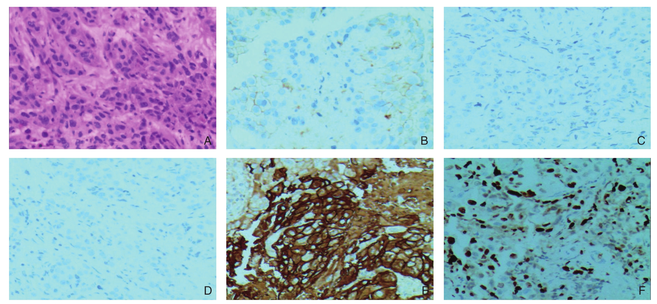
图3 无汇聚征的IDBC肿块的术后病理情况(×400) A:HE染色结果为IDBC;B:Her-2(+);C:ER(-);D:PR(-);E:EGFR(+);F:Ki-67(+)
Figure3 The postoperative pathology of IDBC mass without convergence sign (×400) A: HE staining showing of IDBC; B: Her-2 (+);C: ER (–); D: PR (–); E: EGFR (+); F: Ki-67 (+)
表2 汇聚征与IDBC患者的临床特征的关系[n(%)]
Table2 Relations of the convergence sign with clinicopathologic features of the IDBC patients [n (%)]
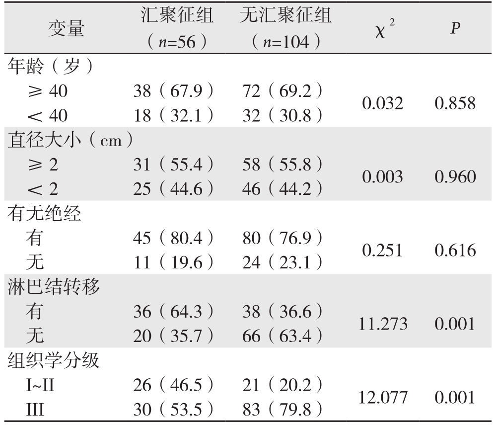
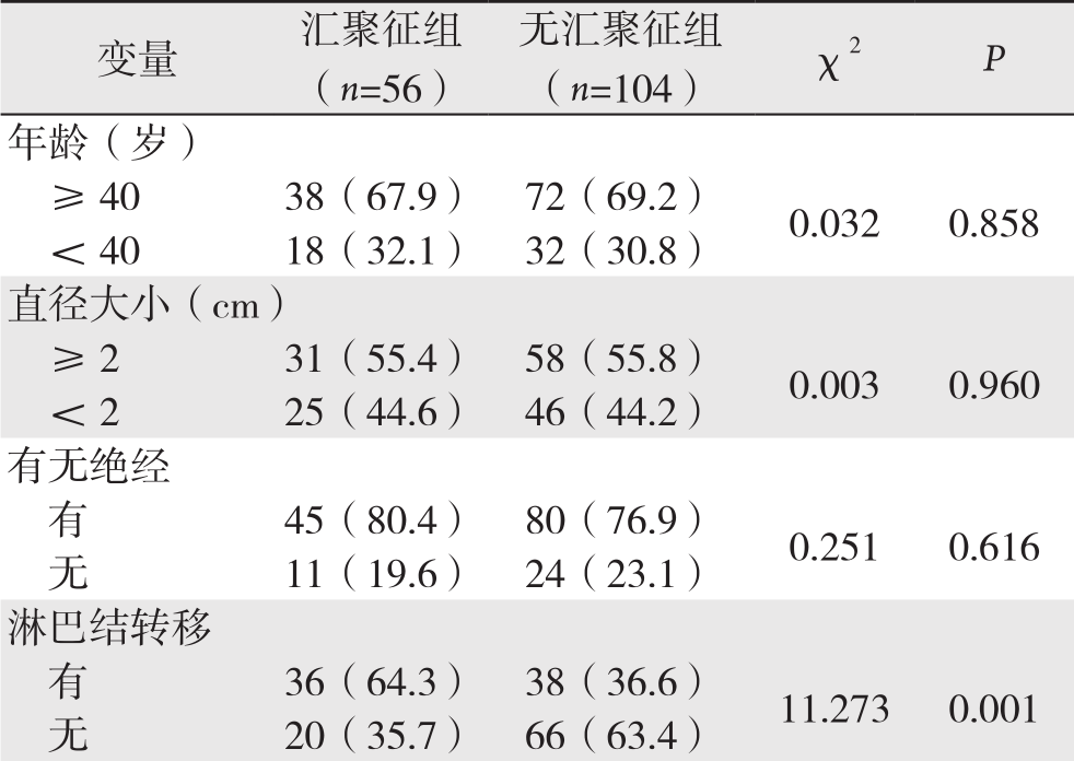 组织学分级I~II 26(46.5) 21(20.2) 12.077 0.001 III 30(53.5) 83(79.8)
3 讨 论
ABVS是一种新的三维容积超声成像系统,通过这一技术可获得整个乳腺连续的横切面,收集这些影像数据,通过计算机自动处理,进行三维重建,可同步获得完整的乳腺矢状面和冠状面的图像。ABVS三维成像技术可形象的显示乳腺肿块与周围腺体组织及乳腺导管的关系,反映肿瘤生长和侵袭改变等生物学信息。乳腺癌超声表现的基础主要是异常的组织病理形态,主要受控制生物学行为特性的基因分子调控。乳腺癌ABVS冠状切面汇聚征表现与分子病理及相关肿瘤基因分子标记物之间的关系研究,有助于进一步明确ABVS冠状切面汇聚征在乳腺癌诊断、预测及治疗中的作用,以期发现新的无创性影像评价指标,用于临床指导乳腺癌患者的个体化精准治疗[13-15]。
表3 IDBC肿块中汇聚征与肿瘤分子标记物的关系[n(%)]
Table3 Relations of the convergence sign of the IDBC masses with tumor markers [n (%)]
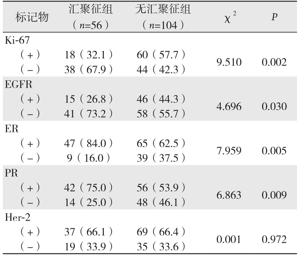
标记物 汇聚征组(n=56)无汇聚征组(n=104) χ2 P Ki-67(+) 18(32.1) 60(57.7) 9.510 0.002(-) 38(67.9) 44(42.3)EGFR(+) 15(26.8) 46(44.3) 4.696 0.030(-) 41(73.2) 58(55.7)ER(+) 47(84.0) 65(62.5) 7.959 0.005(-) 9(16.0) 39(37.5)PR(+) 42(75.0) 56(53.9) 6.863 0.009(-) 14(25.0) 48(46.1)Her-2(+) 37(66.1) 69(66.4) 0.001 0.972(-) 19(33.9) 35(33.6)
随着分子基因技术的飞速发展,近年来乳腺癌相关肿瘤分子标记物日益增多,也逐渐受到人们的关注。目前评价乳腺肿瘤生物学行为及预后最常用的分子标记物主要有ER、PR、Her-2、Ki-67、EGFR等。这些指标可以从多个方面反映乳腺癌的生物特性,指导临床乳腺癌的诊断治疗和预后评估[16-19]。本研究通过分析IDBC肿块在ABVS冠状面的汇聚征与这些分子标记物的关系发现,有汇聚征组Ki-67、EGFR阳性表达率低于无汇聚征组,有汇聚征组ER、PR的阳性表达率均高于无汇聚征组。结果提示汇聚征可在一定程度上反映Ki-67、EGFR、ER、PR的表达水平。当前研究[19-23]显示:Ki-67是增殖细胞核抗原,能反映肿瘤细胞增殖的活性,根据其表达情况,可作为判断肿瘤的恶性程度的指标。患者癌组织中Ki-67高表达,则预示肿瘤恶性程度高,容易侵袭转移,预后差,是一个不良预后的指标。EGFR是一种糖蛋白,主要位于细胞膜,是原癌基因cerbB-1的表达产物,被激活后产生酪氨酸激酶活性,能调节细胞增殖、生长等过程。乳腺癌中EGFR过度表达与肿瘤分化差以及预后不良有关。乳腺癌的生长浸润和扩散转移依赖于体内的激素,尤其与雌、孕激素关系紧密。ER表达阳性在乳腺癌转移中起保护作用,而PR的表达阳性则标志着ER功能的完整。乳腺癌组织中ER、PR受体阴性,则该乳腺癌患者恶性度高,肿瘤分化差,在治疗方面首选化学治疗,内分泌治疗效果不佳。Her-2是一种原癌基因,定位于染色体17q12-21.32上,具有酪氨酸激酶活性。Her-2是评估乳腺癌发生发展的独立预测因子,Her-2阳性表达的乳腺癌患者肿瘤的恶性程度高,容易发生远处转移及术后复发,患者的预后不佳。本研究显示汇聚征与Ki-67、EGFR、ER、PR等肿瘤标记物表达相关,这一结果有助于根据汇聚征这一影像学征象间接反映肿瘤分子标记物的表达水平。无汇聚征的IDBC肿块,多表现为Ki-67阳性、EGFR阳性,这一类患者肿瘤细胞增殖活性强,肿瘤生长快,预后不良。有汇聚征的IDBC肿块,ER阳性或 ER,PR均阳性的发生率高,肿瘤细胞多数分化好,恶性度低,具有更低的侵袭性行为,适合内分泌治疗。ABVS这一影像学技术是一种无创性检查方法,通过观测其冠状面的汇聚特征,可为临床治疗、判断预后提供影像学依据。
本研究发现汇聚征组腋窝淋巴结转移的发生率高于无汇聚征组,汇聚征与腋窝淋巴结转移有关。谭艳娟等[24]、郑逢洋等[25]等也分别在各自的研究中采用多因素逻辑回归分析得出类似结论:淋巴结转移是ABVS冠状切面出现汇聚征的独立危险因素,但其相关机制尚不清楚。淋巴结转移是恶性肿瘤侵袭转移的重要表现之一,但也只是评估乳腺癌生物学行为的一个指标,还需综合考虑其他因素,因此虽然汇聚征与淋巴结转移率呈现出显著的相关性,但仍不足以说明超声声像图上汇聚征征象是乳腺癌强侵袭性的一种表现。Effi等[26]、Liedtke等[27]、Mouttet等[28]的研究均证实乳腺癌患者的年龄、Her-2对分子生物学的表达水平存在一定的影响。在本研究中,直径大小、年龄、Her-2等多个指标与汇聚征作用无明显相关性,这一结果与严丽霞等[29]的研究结果并不一致,这可能与本研究的局限性有关。本研究中样本量不大,纳入的汇聚征相关指标较少,且没有进行多因素逻辑回归分析。因此,关于与IDBC相关的临床病理变量与汇聚征的关系研究,还需纳入其他更多影响预后及治疗的分子生物学及临床指标,以及需要扩大样本量。
综上所述,ABVS作为三维超声成像的新技术,能够为乳腺癌患者提供更多的影像学信息。IDBC肿块在ABVS冠状面呈现的汇聚征与其生物学行为及其调控的肿瘤分子标记物表达密切相关,通过对声像图特征的分析,可以为IDBC肿块的生物学行为及临床精准治疗和预后判断等提供影像学依据。
[1] McPherson K, Steel CM, Dixon JM. ABC of breast diseases. Breast cancer-epidemiology, risk factors, and genetics[J]. BMJ, 2000,321(7261):624–628.
[2] Siegel RL, Miller KD, Jemal A. Cancer Statistics, 2017[J]. CA Cancer J Clin, 2017, 67(1):7–30. doi: 10.3322/caac.21387.
[3] Xu C, Wei S, Xie Y, et al. Three-Dimensional Assessment of Automated Breast Volume Scanner Compared with Handheld Ultrasound in Pre-Operative Breast Invasive Ductal Carcinomas: A Pilot Study of 51 Cases[J]. Ultrasound Med Biol, 2016, 42(9):2089–2096. doi: 10.1016/j.ultrasmedbio.2016.05.007.
[4] Hendriks GA, Hollander B, Menssen J, et al. Automated 3D ultrasound elastography of the breast: a phantom validation study[J].Phys Med Biol, 2016, 61(7):2665–2679. doi: 10.1088/0031–9155/61/7/2665.
[5] Xiao YM, Chen ZH, Zhou QC, et al. The efficacy of automated breast volume scanning over conventional ultrasonography among patients with breast lesions[J]. Int J Gynaecol Obstet, 2015,131(3):293–296. doi: 10.1016/j.ijgo.2015.05.036.
[6] Zheng FY, Yan LX, Huang BJ, et al. Comparison of retraction phenomenon and BI‐RADS‐US descriptors in differentiating benign and malignant breast masses using an automated breast volume scanner[J]. Eur J Radiol, 2015, 84(11):2123–2129. doi: 10.1016/j.ejrad.2015.07.028.
[7] Meng Z, Chen C, Zhu Y, et al. Diagnostic performance of the automated breast volume scanner: a systematic review of inter-rater reliability/agreement and meta-analysis of diagnostic accuracy for differentiating benign and malignant breast lesions[J]. Eur Radiol,2015, 25(12):3638–3647. doi: 10.1007/s00330–015–3759–3.
[8] Kim Y, Kang BJ, Kim SH, et al. Prospective Study Comparing Two Second-Look Ultrasound Techniques: Handheld Ultrasound and an Automated Breast Volume Scanner[J]. J Ultrasound Med, 2016,35(10):2103–2112. doi: 10.7863/ultra.15.11076.
[9] Zheng FY, Yan LX, Huang BJ, et al. Comparison of retraction phenomenon and BI‐RADS‐US descriptors in differentiating benign and malignant breast masses using an automated breast volume scanner[J]. Eur J Radiol, 2015, 84(11):2123–2129. doi: 10.1016/j.ejrad.2015.07.028.
[10] Stuart-Harris R, Caldas C, Pinder SE, et al. Proliferation markers and survival in early breast cancer: a systematic review and meta-analysis of 85 studies in 32,825 patients[J]. Breast, 2008,17(4):323–334. doi: 10.1016/j.breast.2008.02.002.
[11] Soliman NA, Yussif SM. Ki-67 as a prognostic marker according to breast cancer molecular subtype[J]. Cancer Biol Med, 2016,13(4):496–504. doi: 10.20892/j.issn.2095–3941.2016.0066.
[12] Liu Q, Wang XZ, Mu DB, et al. Correlation analysis of hormone receptors and the expressions of HER-2 and Ki-67 in breast cancer[J]. Eur J Gynaecol Oncol, 2015, 36(1):78–83.
[13] Bartlett JM, Starczynski J, Atkey N, et al. HER2 testing in the UK: recommendations for breast and gastric in-situ hybridisation methods[J]. J Clin Pathol, 2011, 64(8):649–653. doi: 10.1136/jcp.2011.089847.
[14] Huang A, Zhu L, Tan Y, et al. Evaluation of automated breast volume scanner for breast conservation surgery in ductal carcinomain situ[J]. Oncol Lett, 2016, 12(4):2481–2484.
[15] Araújo T, Abayazid M, Rutten MJCM, et al. Segmentation and three-dimensional reconstruction of lesions using the automated breast volume scanner (ABVS)[J]. Int J Med Robot, 2017, 13(3).doi: 10.1002/rcs.1767.
[16] Hellgren R, Dickman P, Lei fl and K, et al. Comparison of handheld ultrasound and automated breast ultrasound in women recalled after mammography screening[J]. Acta Radiol, 2017, 58(5):515–520.doi: 10.1177/0284185116665421.
[17] 陈伟财, 何劲松, 宋淑芬, 等. 乳腺癌ER,PR与HER-2,p53的表达及其相关性[J]. 中国普通外科杂志, 2010, 19(8):935–937.Chen WC, He JS, Song SF, et al. Expression of ER, PR, and HER-2, p53 in breast cancer and their correlation[J]. Chinese Journal of General Surgery, 2010, 19(8):935–937.
[18] 高鹏, 周庚寅, 魏军民, 等. 乳腺癌c-erbB2过表达与生存率、内分泌治疗效果和预后的关系[J]. 中国普通外科杂志, 2003,12(10):735–738. doi:10.3969/j.issn.1005–6947.2003.10.006.Gao P, Zhou GY, Wei JM, et al. Relationship of c-erbB2 overexpression with endocrine therapy and prognosis in patients with breast carcinoma[J]. Chinese Journal of General Surgery, 2010,19(8):935–937. doi:10.3969/j.issn.1005–6947.2003.10.006.
[19] 刘新杰, 周文斌, 周冬仙, 等. C-erbB-2,ER及PR表达在乳腺导管内癌与浸润性导管癌的对照研究[J]. 中国普通外科杂志, 2008,17(12):1226–1228.Liu XJ, Zhou WB, Zhou DX, et al. Comparative research on expression of C-erbB-2, ER, PR in ductal carcinoma in situ and infiltrating ductal carcinoma of breast[J]. Chinese Journal of General Surgery, 2008, 17(12):1226–1228.
[20] Elkablawy MA, Albasri AM, Mohammed RA, et al. Ki67 expression in breast cancer. Correlation with prognostic markers and clinicopathological parameters in Saudi patients[J]. Saudi Med J, 2016, 37(2):137–141. doi: 10.15537/smj.2016.2.12285.
[21] Curtis C, Shah S P, Chin SF, et al. The genomic and transcriptomic architecture of 2,000 breast tumours reveals novel subgroups[J].Nature, 2011, 486(7403):346–352. doi: 10.1038/nature10983.
[22] Sinn HP, Schneeweiss A, Keller M, et al. Comparison of immunohistochemistry with PCR for assessment of ER, PR, and Ki-67 and prediction of pathological complete response in breast cancer[J]. BMC Cancer, 2017, 17(1):124. doi: 10.1186/s12885–017–3111–1.
[23] Guo W, Li H, Zhu Y, et al. Prediction of clinical phenotypes in invasive breast carcinomas from the integration of radiomics and genomics data[J]. J Med Imaging (Bellingham), 2015, 2(4):041007.doi: 10.1117/1.JMI.2.4.041007.
[24] 谭艳娟, 包凌云, 黄安茜, 等. 乳腺恶性肿瘤冠状面汇聚征与临床病理学相关因素分析[J]. 中国超声医学杂志, 2015, 31(7):587–589.Tan YJ, Bao LY, Huang AQ, et al. The Relationship between the Retraction Phenomenon on Coronal Plane and Clinical Pathological Characteristics in Breast Carcinoma Patients[J]. Chinese Journal of Ultrasound in Medicine, 2015, 31(7):587–589.
[25] 郑逢洋, 黄备建, 严丽霞, 等. 乳腺癌冠状面汇聚征和生物学行为指标间的相关性研究[J]. 中华超声影像学杂志, 2016, 25(6):496–501. doi:10.3760/cma.j.issn.1004–4477.2016.06.009.Zheng FY, Huang BJ, Yan LX, et al. Correlation between retraction phenomenon on the coronal planes and biological behaviors of breast cancer[J]. Chinese Journal of Ultrasonography, 2016,25(6):496–501. doi:10.3760/cma.j.issn.1004–4477.2016.06.009.
[26] Effi AB, Aman NA, Koui BS, et al. Immunohistochemical determination of estrogen and progesterone receptors in breast cancer: relationship with clinicopathologic factors in 302 patients in Ivory Coast[J]. BMC Cancer, 2017, 17(1):115. doi: 10.1186/s12885–017–3105-z.
[27] Liedtke C, Rody A, Gluz O, et al. The prognostic impact of age in different molecular subtypes of breast cancer[J]. Breast Cancer Res Treat, 2015, 152(3):667–673. doi: 10.1007/s10549–015–3491–3.
[28] Mouttet D, Lae M, Caly M, et al. Estrogen-Receptor, Progesterone-Receptor and HER2 Status Determination in Invasive Breast Cancer.Concordance between Immuno-Histochemistry and MapQuant™Microarray Based Assay[J]. PLoS One, 2016, 11(2):e0146474. doi:10.1371/journal.pone.0146474..
[29] 严丽霞, 黄备建, 刘利民, 等. 汇聚征与乳腺浸润性导管癌临床病理特征的相关性[J]. 中华超声影像学杂志, 2014, 23(12):1057–1060. doi:10.3760/cma.j.issn.1004–4477.2014.12.014.Yan LX, Huang BJ, Liu LM, et al. Correlation between clinicopathological characteristics and convergence symptom in patients with breast in filtrating ductal carcinoma[J]. Chinese Journal of Ultrasonography, 2014, 23(12):1057–1060. doi:10.3760/cma.j.issn.1004–4477.2014.12.014.
