随着腔内技术的发展,主动脉腔内修复术(endovascular aneurysm repair,EVAR)已成为临床治疗腹主动脉瘤(abdominal aortic aneurysms,AAA)的首选治疗方式[1]。研究[2-3]表明,在AAA患者中髂动脉累及的比例为20%~30%。髂动脉分支支架(iliac branched device,IBD)技术作为保留髂内动脉的一种方式,可通过血管内支架置入保留髂动脉的解剖结构和顺向血流。但该技术对髂动脉的长度和直径有一定要求,国内外研究报道仅有部分临床患者适用Cook、Gore的IBD支架,而同时符合两者要求的则更少[4-5]。此外,许多学者研究指出亚洲人群和西方人群在髂动脉解剖中存在显著差异[3,6]。因此,笔者对中国患者髂动脉解剖特点进行分析,进而研究各型IBD支架在中国患者中的适用性。
1 资料与方法
1.1 研究对象
对2015年7月—2017年3月就诊于复旦大学附属中山医院血管外科的AAA患者进行筛选,入组标准:肾下型AAA同时累及单侧或双侧髂动脉,AAA最大直径超过5 cm。髂动脉瘤样扩张直径超过正常值的1.5倍及以上;中国人群髂总动脉直径正常指标表明,男性为11~12 mm,女性为10~11 mm。排除标准:动脉瘤破裂、假性动脉瘤或霉菌性动脉瘤患者。
1.2 研究方法
收集每名患者术前计算机断层扫描血管造影(computed tomography angiography,CTA)及三维重建资料。应用Vitrea fX software(Vital Images,Minnetonka,MN,USA)3D工作站,对AAA最大径、髂动脉及其分支的长度和最大径等数据进行测量,分别计算中位数、平均数和标准差。所有数据的收集和计算由2名研究人员分别独立完成。所有数据的汇总、计算和比较在Excel(Microsoft,Redmond,WA,USA)和SPSS(IBM Corp,Armonk,NY,USA)软件中进行。根据Cook公司的Zenith IBD和GORE公司的Excluder IBE两款IBD支架的使用标准(表1),以及产品说明书,分析中国患者病变髂动脉的适用性。
表1 两种支架的解剖排除标准
Table 1 Anatomic exclusion criteria for the two types of stents
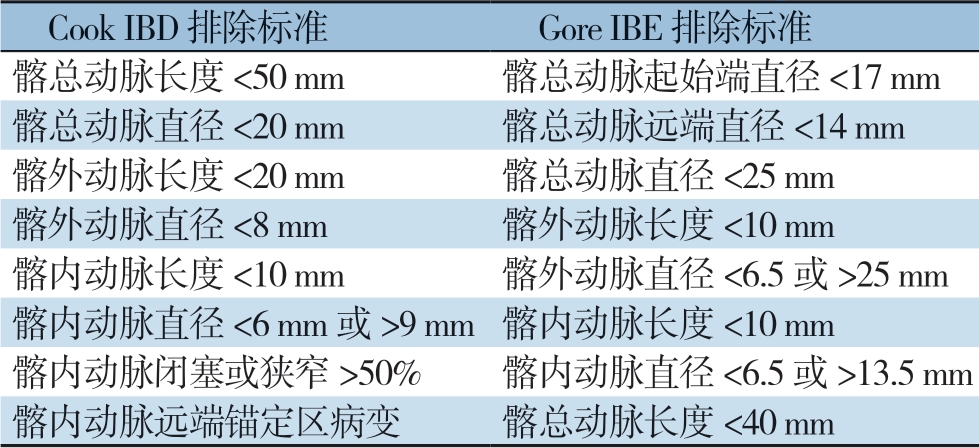
Cook IBD 排除标准 Gore IBE 排除标准髂总动脉长度<50 mm髂总动脉起始端直径<17 mm髂总动脉直径<20 mm髂总动脉远端直径<14 mm髂外动脉长度<20 mm髂总动脉直径<25 mm髂外动脉直径<8 mm 髂外动脉长度<10 mm髂内动脉长度<10 mm髂外动脉直径<6.5或>25 mm髂内动脉直径<6 mm或>9 mm髂内动脉长度<10 mm髂内动脉闭塞或狭窄>50% 髂内动脉直径<6.5或>13.5 mm髂内动脉远端锚定区病变髂总动脉长度<40 mm
1.3 数据处理
计量指标等采用均数±标准差( ±s)或中位数(范围)[M(范围)]表示,计数指标用例数(百分比)[n(%)]表示。
2 结 果
2.1 基本情况及相关数据
共有58例病变累及髂总动脉的肾下型AAA患者入组,男51例,女7例;平均年龄(73.5±7.7)岁。其中累及双侧髂总动脉49例,单侧9例,累及髂动脉总数107支。AAA瘤体的平均长度为(103.3±32.3)mm;左右髂总动脉长度相似,分别为(57.9±18.1)mm和(56.7±17.4)mm。然而,与左侧髂总动脉直径(17.7±7.2)mm相比,右侧髂总动脉直径则较大(25.1±9.4)mm。两侧同时受病变累及的患者髂内、髂外动脉长度和直径相似(表2)。
2.2 适用性及限制因素分析
本研究中107支病变髂动脉对两种IBD支架的适用性结果见表3。对于Cook IBD最主要的限制是髂内动脉直径>9 mm或者<6 mm(n=50,46.7%),以及髂总动脉直径<20 mm(n=36,33.6%);Gore IBE最主要的限制是髂内动脉直径>13.5 mm或者<6.5 mm(n=29,27.1%),以及髂总动脉直径<25 mm(n=67,62.6%)。统计结果表明两种支架的解剖适用率分别为26.1%(28/107,Cook IBD)和20.6%(22/107,Gore IBE)。接近一半的病变血管42.9%(46/107)因单个解剖因素受限而无法使用Cook IBD支架;近1/4的病变血管23.5%(25/107)受两种解剖因素限制。而对于Gore IBE受单一和任意两种解剖因素限制的比例相差较大,分别为58.8%(63/107)和16.9%(18/107)(表4)。
表2 AAA累及髂动脉的相关解剖测量和数据分析(mm)
Table 2 Relevant anatomic measurements and data analysis of AAA involving the iliac arteries (mm)
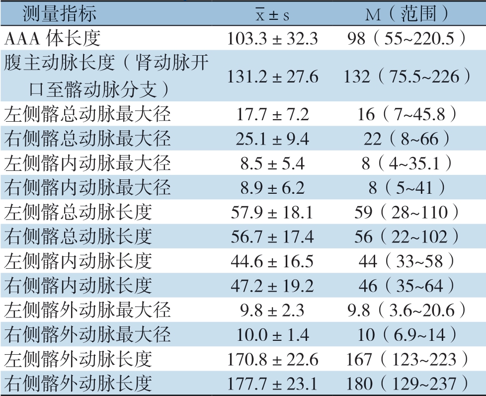
测量指标images/BZ_15_1000_2138_1036_2179.png±sM(范围)AAA体长度103.3±32.398(55~220.5)腹主动脉长度(肾动脉开口至髂动脉分支)131.2±27.6132(75.5~226)左侧髂总动脉最大径17.7±7.216(7~45.8)右侧髂总动脉最大径25.1±9.422(8~66)左侧髂内动脉最大径8.5±5.48(4~35.1)右侧髂内动脉最大径8.9±6.28(5~41)左侧髂总动脉长度57.9±18.159(28~110)右侧髂总动脉长度56.7±17.456(22~102)左侧髂内动脉长度44.6±16.544(33~58)右侧髂内动脉长度47.2±19.246(35~64)左侧髂外动脉最大径9.8±2.39.8(3.6~20.6)右侧髂外动脉最大径10.0±1.410(6.9~14)左侧髂外动脉长度170.8±22.6167(123~223)右侧髂外动脉长度177.7±23.1180(129~237)
表3 两种支架在107支病变血管中的适用性[n(%)]
Table 3 Applicability of the two types of stents in the107 diseased vessels [n (%)]
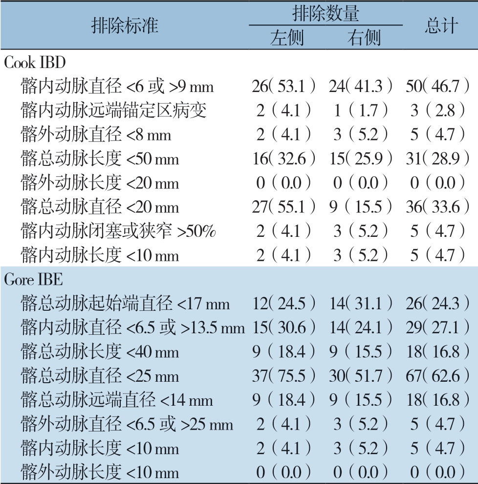
排除标准排除数量总计左侧右侧Cook IBD 髂内动脉直径<6或>9 mm26(53.1)24(41.3)50(46.7) 髂内动脉远端锚定区病变2(4.1)1(1.7)3(2.8) 髂外动脉直径<8 mm2(4.1)3(5.2)5(4.7) 髂总动脉长度<50 mm16(32.6)15(25.9)31(28.9) 髂外动脉长度<20 mm0(0.0)0(0.0)0(0.0) 髂总动脉直径<20 mm27(55.1)9(15.5)36(33.6) 髂内动脉闭塞或狭窄>50%2(4.1)3(5.2)5(4.7) 髂内动脉长度<10 mm2(4.1)3(5.2)5(4.7)Gore IBE 髂总动脉起始端直径<17 mm12(24.5)14(31.1)26(24.3) 髂内动脉直径<6.5或>13.5 mm15(30.6)14(24.1)29(27.1) 髂总动脉长度<40 mm9(18.4)9(15.5)18(16.8) 髂总动脉直径<25 mm37(75.5)30(51.7)67(62.6) 髂总动脉远端直径<14 mm9(18.4)9(15.5)18(16.8) 髂外动脉直径<6.5或>25 mm2(4.1)3(5.2)5(4.7) 髂内动脉长度<10 mm2(4.1)3(5.2)5(4.7) 髂外动脉长度<10 mm0(0.0)0(0.0)0(0.0)
表4 两种支架的总体解剖受限因素[n(%)]
Table 4 The anatomic restricting factors for the two types of stents [n (%)]
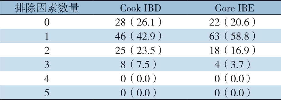
排除因素数量Cook IBDGore IBE 0 28(26.1)22(20.6)1 46(42.9)63(58.8)2 25(23.5)18(16.9)3 8(7.5)4(3.7)4 0(0.0)0(0.0)5 0(0.0)0(0.0)
3 讨 论
标准的EVAR手术需要足够的远端和近端锚定区,当AAA累及单侧或双侧髂总动脉时,约有1/3的患者因锚定区不足需要同时治疗髂动脉病变[7]。髂动脉瘤的成功治疗一方面是髂外及髂内动脉的血流通畅,另一方面是对髂总动脉的长期保护[8-9]。为确保有足够的锚定区,常见的方法是把髂总动脉内的支架远端延长至髂外动脉,同时伴或不伴髂内动脉的栓塞和覆盖[10-12]。然而,对于髂总动脉病变的患者,确保至少一侧髂内动脉血流通畅能有效的避免相关并发症,改善患者的生活质量[13-15]。 若下腹部动脉尤其是双侧髂内动脉同时被覆盖时,可能造成严重的术后并发症包括臀肌缺血跛行(28%~42%),阳痿(17%~24%),结肠缺血(3.4%)和脊髓缺血(0.1%~0.3%)[16-18]。研究[14]还显示,单侧髂内动脉闭塞臀部缺血跛行率为28%(198/706);双侧髂内动脉闭塞臀部缺血跛行率为42%(43/102)。2009年,Verzini等[19]对Cook IBD支架保留髂内动脉研究结果提示:相比于对照组,保留髂内动脉的实验组其支架内漏(4% vs. 19%)和臀部缺血跛行(4% vs. 22%)发生率都较低。他们的结论是,年轻且髂动脉解剖满足条件的患者,应积极进行保留髂内动脉的治疗。一些研究已经证实,IBD之外的髂内动脉保留技术也能有效的降低术后并发症机率,包括外科手术、杂交手术、三明治技术、髂外动脉-髂内动脉腔内分流技术以及弹簧栓塞技术[7,11,20-24]。2001年至今,经历十几年的发展,IBD技术的广泛应用得到了大量临床实践和长期术后随访的论证支持[25];但是由于不同种族人群解剖差异,需具体分析该技术在不同情况中的适用性。
自肾动脉平面以下至髂动脉分叉处,相比于西方人群(183.6±28.3)mm,中国患者(131.2±27.6)mm长度较短,可能原因是中国人群的平均身高相对较低[26-27]。对病变髂动脉的解剖特点分析:髂总动脉左侧平均长度57.9 mm,右侧平均长度56.7 mm,与之前日本学者Itoga等[28]报道的AAA患者双侧髂总动脉平均长度56.5 mm相近;但是短于西方患者双侧髂总动脉平均长度70.8~72 mm[10]。研究发现与左侧(17.7±7.2)mm相比,右侧髂总动脉直径(25.1±9.4)mm受累扩张更明显,但仍小于美国患者左侧髂总最大直径27.1 mm;右侧髂总最大直径31.7 mm[10]。西方学者、日本学者以及本次研究对于两种IBD解剖限制性的对比更加直观的见于图1。髂总动脉直径是限制IBD使用的关键因素之一,Gore IBE的排除比率为62.6%(67/107)。而对于Cook IBD髂内动脉的的直径则是最主要的限制因素,本次研究的排除比率为46.7%(50/107)。本研究结果显示,病变患者左右髂内动脉和髂外动脉相比较,其直径和长度是相似的。我国髂动脉病变患者和西方患者在解剖学上存在显著差异,系统的分析中国患者髂动脉解剖特点是必要的。
Gore IBE要求髂总动脉的近端开口直径>17 mm以便于支架的完全打开,Cook IBD则没有单独的髂动脉近端开口要求[29]。髂内动脉对锚定区直径的要求是两款髂动脉分支支架使用受限的最主要原因。Cook IBD 髂内动脉直径的解剖要求是6~9 mm,排除了46.7%的病变血管。这一缺陷在2014年开始的新版本Cook IBD试验中得到了一些改进,对于本次研究6~10 mm的要求将使受限比率从46.7%降低到38.5%[30]。而对于Gore IBE髂内动脉直径的解剖要求是一个更广泛的治疗范围6.5~13.5 mm,但依然有27.1%的排除率 。为了更好在临床工作中开展IBD技术,下一代产品的开发应着重于主要的解剖限制因素的解决。除外5例因病变完全闭塞的髂内动脉,剩余全部满足两种支架的长度要求。对于IBD技术的适用性比率,本研究的结论与国内外已报道的结果一致。
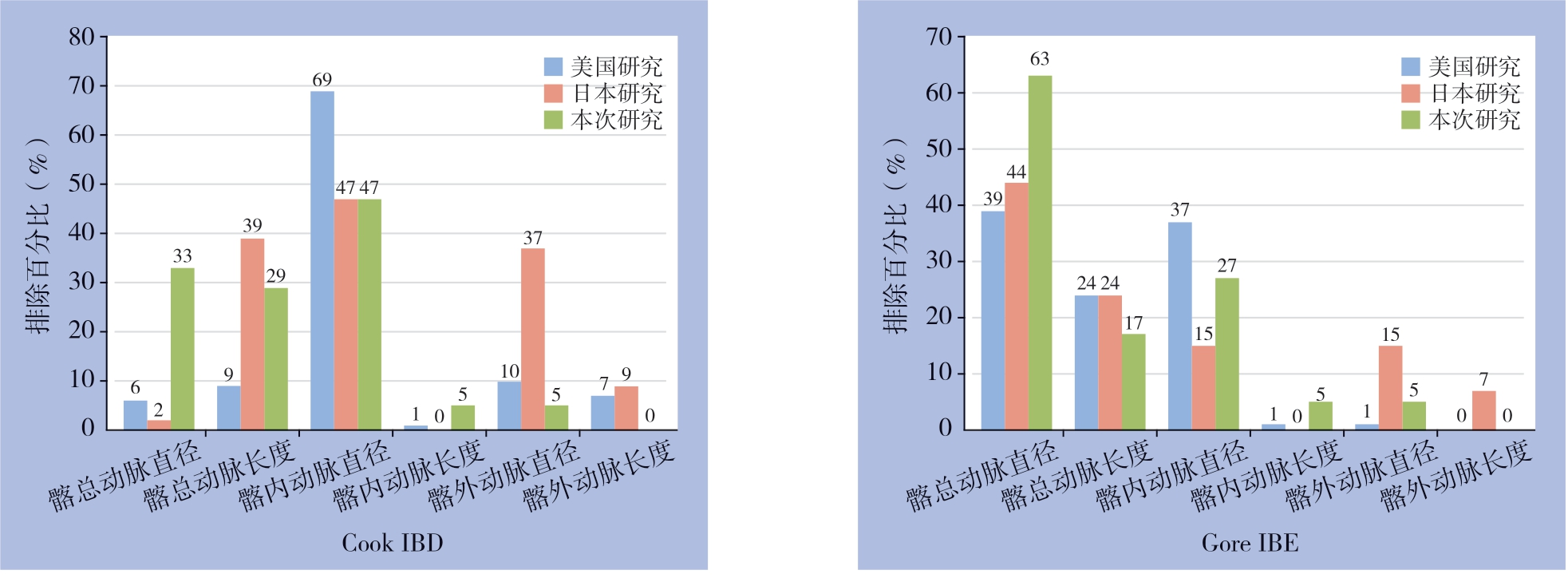
图1 3项研究中不同IBD解剖限制条件的排除百分比
Figure 1 Exclusion percentages of different IBD restricting features in the three studies.
本组研究有一定的局限性:一是回顾性调查研究,并且相对总体的AAA患者入组的累及髂动脉病变的患者数量较少;二是入组患者的解剖学评估完全依靠术前影像学资料;三是其他因素例如心脏功能、肾功能衰竭、药物使用等对数据结果的影响。
通过解剖特点的分析证实相比于西方人群,中国患者的病变髂总动脉直径较小、长度较短。中国和欧美患者对两种IBD支架的适用性存在一定差异。
[1]Chaikof EL, Dalman RL, Eskandari MK, et al. The Society for Vascular Surgery practice guidelines on the care of patients with an abdominal aortic aneurysm[J]. J Vasc Surg, 2018, 67(1): 2-77. doi: 10.1016/j.jvs.2017.10.044.
[2]Henretta JP, Karch LA, Hodgson KJ, et al. Special iliac artery considerations during aneurysm endografting[J]. Am J Surg, 1999, 178(3):212-218.
[3]Cheng SW, Ting AC, Ho P, et al. Aortic aneurysm morphology in Asians: features affecting stent-graft application and design[J]. J Endovasc Ther, 2004, 11(6):605-612. doi: 10.1583/04-1268R.1.
[4]Loth AG, Rouhani G, Gafoor SA, et al. Treatment of iliac artery bifurcation aneurysms with the second-generation straight iliac bifurcated device[J]. J Vasc Surg, 2015, 62(5):1168-1175. doi: 10.1016/j.jvs.2015.06.135.
[5]Marques de Marino P, Botos B, Kouvelos G, et al. Use of Bilateral Cook Zenith Iliac Branch Devices to Preserve Internal Iliac Artery Flow During Endovascular Aneurysm Repair[J]. Eur J Vasc Endovasc Surg, 2019, 57(2):213-219. doi: 10.1016/j.ejvs.2018.08.002.
[6]Midorikawa H, Ogawa T, Satou K, et al. Morphological study of abdominal aortic aneurysm: optimal stent-graft size for Japanese patients[J]. Ann Thorac Cardiovasc Surg, 2006, 12(2):121-125.
[7]Youn JK, Kim SM, Han A, et al. Surgical Treatment of Infected Aortoiliac Aneurysm[J]. Vasc Specialist Int, 2015, 31(2):41-46. doi: 10.5758/vsi.2015.31.2.41.
[8]赵珺. 腹主动脉瘤腔内修复术中髂内动脉的疏与堵[J]. 中国普通外科杂志, 2017, 26(12):1516-1524. doi:10.3978/j.issn.1005-6947.2017.12.002. Zhao J. Preservation or occlusion of the hypogastric artery in endovascular abdominal aortic aneurysm repair[J]. Chinese Journal of General Surgery, 2017, 26(12):1516-1524. doi:10.3978/j.issn.1005-6947.2017.12.002.
[9]Schneider DB, Matsumura JS, Lee JT, et al. Prospective, multicenter study of endovascular repair of aortoiliac and iliac aneurysms using the Gore Iliac Branch Endoprosthesis[J]. J Vasc Surg, 2017, 66(3):775-785. doi: 10.1016/j.jvs.2017.02.041.
[10]Pearce BJ, Varu VN, Glocker R, et al. Anatomic suitability of aortoiliac aneurysms for next generation branched systems[J]. Ann Vasc Surg, 2015, 29(1):69-75. doi: 10.1016/j.avsg.2014.08.003.
[11]Naughton PA, Park MS, Kheirelseid EA, et al. A comparative study of the bell-bottom technique vs hypogastric exclusion for the treatment of aneurysmal extension to the iliac bifurcation[J]. J Vasc Surg, 2012, 55(4):956-962. doi: 10.1016/j.jvs.2011.10.121.
[12]谷涌泉, 刘一人, 郭连瑞, 等. 主动脉腔内修复术中髂内动脉保留技术[J]. 介入放射学杂志, 2017, 26(2):184-187. doi:10.3969/j.issn.1008-794X.2017.02.021.Gu YQ, Liu YR, Guo LR, et al. Preservation technique of internal iliac artery in performing endovascular aortic repair[J]. Journal of Interventional Radiology, 2017, 26(2):184-187. doi:10.3969/j.issn.1008-794X.2017.02.021.
[13]何玉祥, 种振岳, 王默, 等. 腹主动脉瘤腔内隔绝术中髂动脉的处理[J]. 外科理论与实践, 2011, 16(2):133-136.He YX, Zhong ZY, Wang M, et al. Management of iliac arteries in endovascular graft exclusion of abdominal aortic aneurysm[J]. Journal of Surgery Concepts & Practice, 2011, 16(2):133-136.
[14]Farahmand P, Becquemin J P, Desgranges P, et al. Is hypogastric artery embolization during endovascular aortoiliac aneurysm repair (EVAR) innocuous and useful?[J]. Eur J Vasc Endovasc Surg, 2008, 35(4):429-435. doi: 10.1016/j.ejvs.2007.12.001.
[15]Schneider DB, Milner R, Heyligers JMM, et al. Outcomes of the GORE Iliac Branch Endoprosthesis in clinical trial and realworld registry settings[J]. J Vasc Surg, 2019, 69(2):367-377. . doi: 10.1016/j.jvs.2018.05.200.
[16]陈宏宇, 戴贻权, 郭平凡. EVAR中保留髂内动脉的腔内手术技术[J]. 血管与腔内血管外科杂志, 2015, 1(1):27-31.Chen HY, Dai YQ, Guo PF. Techniques for presevation of internal iliac artery during EVAR[J]. Journal of Vascular and Endovascular Surgery, 2015, 1(1):27-31.
[17]Lin PH, Chen AY, Vij A. Hypogastric artery preservation during endovascular aortic aneurysm repair: is it important?[J]. Semin Vasc Surg, 2009, 22(3):193-200. doi: 10.1053/j.semvascsurg.2009.07.012.
[18]Freyrie A, Testi G, Gargiulo M, et al. Spinal cord ischemia after endovascular treatment of infrarenal aortic aneurysm. Case report and literature review[J]. J Cardiovasc Surg (Torino), 2011, 52(5):731-734.
[19]Verzini F, Parlani G, Romano L, et al. Endovascular treatment of iliac aneurysm: Concurrent comparison of side branch endograft versus hypogastric exclusion[J]. J Vasc Surg, 2009, 49(5):1154-1161. doi: 10.1016/j.jvs.2008.11.100.
[20]郭俊莹, 戴向晨, 朱杰昌, 等. 髂动脉分支支架治疗髂动脉瘤的近期结果分析[J]. 血管与腔内血管外科杂志, 2017, 3(6):1065-1068. doi:10.19418/j.cnki.issn2096-0646.2017.06.11.Guo JY, Dai XC, Zhu JC, et al. Short-term results of iliac artery branch device in the treatment of iliac artery aneurysm[J]. Journal of Vascular and Endovascular Surgery, 2017, 3(6):1065-1068. doi:10.19418/j.cnki.issn2096-0646.2017.06.11.
[21]韩晓峰, 黄小勇, 郭曦, 等. GORE EXCLUDER覆膜支架在腹主动脉瘤腔内修复术中的临床应用[J]. 心肺血管病杂志, 2013, 32(6):714-716. doi:10.3969/j.issn.1007-5062.2013.06.016.Han XF, Huang XY, Guo X, et al. Endovascular repair of abdominal aortic aneurysm with GORE EXCLUDER stent grafts[J]. Journal of Cardiovascular and Pulmonary Diseases, 2013, 32(6):714-716. doi:10.3969/j.issn.1007-5062.2013.06.016.
[22]Lobato AC. Sandwich technique for aortoiliac aneurysms extending to the internal iliac artery or isolated common/internal iliac artery aneurysms: a new endovascular approach to preserve pelvic circulation [J]. J Endovasc Ther, 2011, 18(1):106-111. doi: 10.1583/10-3320.1.
[23]DeRubertis BG, Quinones-Baldrich WJ, Greenberg JI, et al. Results of a double-barrel technique with commercially available devices for hypogastric preservation during aortoilac endovascular abdominal aortic aneurysm repair[J]. J Vasc Surg, 2012, 56(5):1252-1259. doi: 10.1016/j.jvs.2012.04.070.
[24]Pirkl M, Krampota F, Daněk T, et al. Early surgical and vascular complications after open repairs of aortoiliac aneurysms [J]. Rozhl Chir, 2018, 97(11):499-503.
[25]Jongsma H, Bekken JA, Bekkers WJ, et al. Endovascular Treatment of Common Iliac Artery Aneurysms With an Iliac Branch Device: Multicenter Experience of 140 Patients[J]. J Endovasc Ther, 2017, 24(2):239-245. doi: 10.1177/1526602816679132.
[26]Fryar CD, Gu Q, Ogden CL, et al. Anthropometric Reference Data for Children and Adults: United States, 2011-2014[J]. Vital Health Stat 3, 2016, (39):1-46.
[27]Fryar CD, Gu Q, Ogden CL. Anthropometric reference data for children and adults: United States, 2007-2010[J]. Vital Health Stat 11, 2012, (252):1-48.
[28]Itoga NK, Fujimura N, Hayashi K, et al. Outcomes of Endovascular Repair of Aortoiliac Aneurysms and Analyses of Anatomic Suitability for Internal Iliac Artery Preserving Devices in Japanese Patients[J]. Circ J, 2017, 81(5):682-688. doi: 10.1253/circj.CJ-16-1109.
[29]Millon A, Della Schiava N, Arsicot M, et al. Preliminary Experience with the GORE((R)) EXCLUDER((R)) Iliac Branch Endoprosthesis for Common Iliac Aneurysm Endovascular Treatment[J]. Ann Vasc Surg, 2016, 33:11-17. doi: 10.1016/j.avsg.2015.12.003.
[30]Li Y, Hu Z, Zhang J, et al. Iliac Aneurysms Treated with Endovascular Iliac Branch Device: A Systematic Review and Meta-analysis[J]. Ann Vasc Surg, 2019, 56:303-316. doi: 10.1016/j.avsg.2018.07.058.
