肝细胞癌(hepatocellular carcinoma,HCC)是世界上第五大常见的恶性肿瘤,其病死率在我国高居第二位[1]。目前HCC的治疗方式主要有手术切除、肝移植、经导管肝动脉化疗栓塞(transarterial chemoembolization,TACE)、局部消融以及以索拉菲尼为代表的药物治疗等[2]。肝移植是虽然被认为是HCC潜在的根治性治疗方案,但供体短缺和费用高昂限制了肝移植的推广。TACE和局部消融在预后方面并不令人满意且适应证较为局限[3]。免疫治疗和靶向药物治疗虽取得了一些成果但尚未得到广泛探索[4]。所以手术切除仍然是目前治疗HCC的最有效和最常用的手段之一[5-6],主要包括解剖性切除(anatomic resection,AR)和非解剖性切除(non-anatomic resection,NAR)。
然而,多年来关于AR和NAR治疗HCC的争议不断。争议的核心焦点是两种术式的彻底性与安全性[5]。有学者[7-9]认为AR能最大程度地切除肿瘤,减少复发风险,因而倾向于选择AR。也有研究[10-11]表明NAR在手术安全性方面更具优势,保留足够肝组织对患者预后更重要。而坚持两种术式疗效无差异的研究[12-13]也不少。HCC手术切除后的高复发率和转移率是长期困扰临床医生、阻碍肝脏外科发展的难题,因此能否降低术后复发和转移也成为AR和NAR对比研究的重要成果之一[5-6]。此外,微血管侵犯(microvascular invasion,MVI)与HCC早期复发存在明显相关性[9,14],合并MVI的HCC有一定特殊性,预后更差,在手术切除方式的选择上也争议较大。
鉴于以上临床中亟待解决的问题,本研究通过纳入最新的随机对照试验[1]和队列研究[7,10-13,15-41],系统评价AR和NAR治疗HCC患者和合并MVI的HCC患者的近期及远期疗效,得出可靠结论,为临床实践提供理论指导。
1 资料与方法
1.1 纳入与排除标准
1.1.1 纳入标准 ⑴研究类型:随机对照试验(randomized controlled trial,RCT)或队列研究,无语种限制。⑵研究对象:接受AR 或NAR 的HCC 患者,其性别、年龄、种族不限。⑶干预措施:试验组采用AR 进行手术,对照组采用NAR 进行手术。⑷结局指标:主要结局指标包括:早期复发率、肝内局部复发率、肝内远处复发率、肝外转移发生率、无瘤生存率(disease-free survival,DFS)、总生存率(overall survival,OS);次要结局指标包括:手术时间、术中出血量、输血率、术后并发症发生率、住院时间。
1.1.2 排除标准 ⑴二次手术或转移性HCC 患者。⑵无法提取结局指标等有效数据的研究。⑶动物实验、综述、Meta 分析、病例报道、摘要、会议记录或专家意见等。⑷重复发表的文献。
1.2 文献检索策略
计算机检索Webof Science、PuMmed、Embase、Cochrane Library、CBM、WanFang Data和CNKI数据库,检索比较AR与NAR在治疗HCC疗效的临床研究,检索时限均为建库至2020年9月。采用主题词与自由词结合的检索方式进行文献检索。中文检索词:肝癌;肝细胞癌;肝脏恶性肿瘤;解剖性切除;非解剖性切除;肝切除术。英文检索词:Hepatocellular Carcinoma;LiverCell Carcinoma;Liver Neoplasms;Anatomic Resection;Non-anatomic Resection;Hepatectomy。
1.3 文献筛选与数据提取
由2 名研究者独立筛选文献、提取资料并交叉核对。如遇分歧,则通过讨论或与第三方协商解决。文献筛选时首先阅读文题,在排除明显不相关的文献后,进一步阅读摘要和全文以确定是否纳入。资料提取内容包括:⑴纳入研究的基本信息:第一作者姓名、研究类型、发表年份等;⑵研究对象的基线特征和干预措施;⑶偏倚风险评价的关键要素;⑷所关注的结局指标和结果测量数据。
1.4 文献质量评价
由2 名研究者独立评价纳入文献的偏倚风险并交叉核对结果。对于队列研究,采用纽卡斯尔-渥太华量表(The Newcastle-Ottawa Scale,NOS)[42]评价偏倚风险,总分为9分,≥7分为高质量文献,纳入文献要求≥6 分;对于RCT,采用Cochrane 手册5.1.0推荐的RCT偏倚风险评估工具[43]评价偏倚风险。
1.5 统计学处理
采用RevMan 5.3软件进行Meta分析。计量资料采用均数差(mean difference,MD)为效应分析统计量,二分类变量采用比值比(odds ratio,OR)为效应分析统计量,各效应量均计算其95% CI。通过I2定量判断研究间的异质性大小。若各研究间无明显异质性(I2<50%,P>0.10),则采用固定效应模型进行Meta分析;若各研究结果间存在明显异质性(I2>50%,P<0.10),进一步分析异质性来源,并采用亚组分析或敏感度分析等方法进行处理,若异质性仍无法消除,则采用随机效应模型进行Meta分析。
2 结果
2.1 文献筛选流程及结果
初检共获得相关文献2251篇,经逐层筛选,最终纳入33个研究,包括1个RCT[1]和32个队列研究[7,10-13,15-41],共6132例患者,其中AR组3029例,NAR组3103例。具体文献筛选流程及结果见图1。

图1 文献筛选流程及结果
Figure 1 Literature screening process and results
2.2 纳入研究的基本特征及偏倚风险评价结果
纳入研究的基本特征见表1。队列研究的偏倚风险评价结果见表2;RCT的偏倚风险评价结果见表3。
2.3 Meta 分析结果
2.3.1 早期复发率 共12 个研究报道了早期复发率[1,12-13,15,19-20,22,24,32,37-38,41]。异质性检验显示各研究间有异质性(P=0.0002,I2=69%),采用随机效应模型。Meta 分析结果显示:与NAR 比较,AR 能减少患者术后早期复发率(OR=0.67,95% CI=0.49~0.92,P=0.01)(图2)。
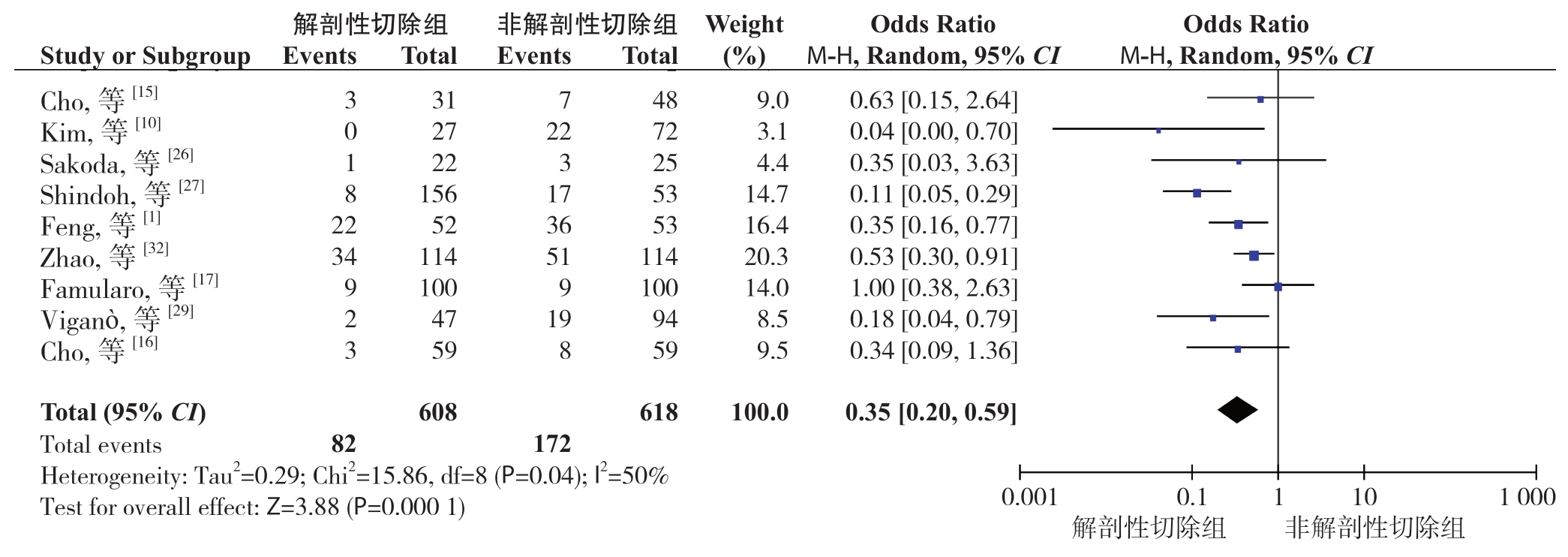
2.3.2 肝内局部复发率 共9 个研究报道了肝内局部复发率[1,10,15-17,26-27,29,32]。异质性检验显示各研究间有异质性(P=0.04,I2=50%),采用随机效应模型。Meta 分析结果显示:与NAR 比较,AR 能明显减少患者术后肝内局部复发率(OR=0.35,95% CI=0.20~0.59,P=0.0001)(图3)。
表1 纳入研究的基本特征
Table 1 General characteristics of the included studies

注:1)早期复发率;2)肝内局部复发率;3)肝内远处复发率;4)肝外转移发生率;5)1年DFS;6)2年DFS;7)3年DFS;8)5年DFS;9)1年OS;10)2年OS;11)3年OS;12)5年OS
Note:1) Early recurrence rate;2) Intrahepatic local recurrence rate;3) Intrahepatic distant recurrence rate;4) Extrahepatic metastasis rate;5) 1-year DFS;6) 2-year DFS;7) 3-year DFS;8) 5-year DFS;9) 1-year OS;10) 2-year OS;11) 3-year OS;12) 5-year OS
主要结局指标5),7),8),9),11),12)1),4),5),6),7),8),9),10),11),12)5),6),7),8),9),10),11),12)5),7),8),9),11),12)5),6),7),8),9),10),11),12)1),4),5),6),7),8),9),10),11),12)2),5),6),7),8),9),10),11),12)2),3),4),5),7),8),9),11),12)2),3),4),5),6),7),8),9),10),11),12)1),2),3),4),5),6),7),8),9),10),11),12)4),7),11),5),6),7),9),10),11),12)1),4),5),6),7),8),9),10),11),12)1),2),3),4),5),6),7),8),9),10),11),12)5),6),7),8),9),10),11),12)1),2),3),4),5),6),7),8),9),10),11),12)1),4),5),6),7),9),10),11),5),6),7),8),9),10),11),12)5),6),7),8),9),10),11),12)2),3),4),5),6),7),8),1),5),6),7),8),9),10),11),12)5),6),7),9),10),11),2),5),6),7),8),9),10),11),12)1),5),6),9),10),4),5),6),7),9),11),12)1),5),6),7),8),9),10),11),12)5),6),7),8),9),10),11),12)2),3),4),5),6),7),8),9),10),11),12)1),5),6),7),8),9),10),11),12)5),6),7),8),9),10),11),12)4),5),6),7),9),10),11),1),4),5),6),7),8),9),10),11),12)5),6),7),8),9),10),11),12)22257032106260244841137331127613035647503612510963154741349肝硬化(n)试验组 对照组1226未报道 未报道29559951未报道 未报道063926123296075136504未报道 未报道93未报道 未报道2525747443160771147未报道 未报道对照组66/664/0 Child 分级(A/B,n)53/2093/3未报道B A 或53/021/472/048/057/6 B A 或350/553/0141/0113/1 B A 或40/819/184/1347/037/1294/046/1170/017/0185/059/0未报道103/051/0110/480/661/3试验组67/5131/1242/2未报道B A 或156/022/027/031/058/0 B A 或353/252/095/0112/2 B A 或116/919/185/1493/031/447/042/0100/017/091/059/0未报道103/051/0114/082/460=91/9456±866±8对照组65.5±9.463.1±8.357.9±10.850.8±13.1未报道49.2±13.060.1±9.859.0±7.148.1±11.260.5±8.854.9±10.767(39~83)67(30~86)49(27~6)59.5(42~77)65(16~78)68(41~79)50(28~70)64(27~86)49(27~75)57(30~81)74(49~84)59(32~73)69.5(23~85)56(23~84)57(30~73)67(48~77)68.5(39~86)年龄(岁)试验组71(44~83)69(32~84)66.4±8.849(27~76)58.5(20~80)63.2±8.467(13~85)72(53~81)51(24~78)58.9±11.653±1151.7±9.765(28~85)42(18~66)未报道55(29~82)48.8±12.367±1073(31~82)67.5(59.2~74.5)66.6(58.9~73.2)63(28~70)61.7±8.771(48~81)57.4±10.546.9±11.561.4±10.1<60/≥60=54/37<60/≥54.9±11.557(31~82)53(29~82)51.0(25.0~80.0)54.0(26.0~80.0)67(48~78)69.5(42~84)/女,n)对照组51/1358/1457/1686/1034/9257/7239/1419/656/1632/1651/1238/7272/8347/6131/1094/20151/2629/1915/529/7129/1842/775/1939/8156/142/15160/2545/1479/1184/1941/1088/2667/19样本量(男试验组55/952/20113/3036/865/23260/69122/3412/1021/624/742/1625/4274/8144/879/1693/21148/29106/1916/429/7161/3229/638/933/988/122/1578/1343/1676/1484/1939/1290/2465/21研究类型队列研究队列研究队列研究队列研究队列研究队列研究队列研究队列研究队列研究队列研究队列研究队列研究队列研究RCT队列研究队列研究队列研究队列研究队列研究队列研究队列研究队列研究队列研究队列研究队列研究队列研究队列研究队列研究队列研究队列研究队列研究队列研究队列研究国家日本日本日本中国韩国日本日本日本韩国韩国中国中国日本中国中国中国中国日本日本意大利中国中国意大利中国中国中国韩国中国中国中国日本日本[23]2015[24]2019 西班牙作者及年份[25]2014[13]2015[11]2015[12]2015[39]2016[40]2017[31]2017[17]2018[37]2018[41]2019[38]2019 Okamura,等Hirokawa,等Matsumoto,等[27]2016,等[21]2015[26]2016[10]2016[15]2016[36]2016,等[19]2017[1]2017,等[32]2017[7]2018,等[35]2018[29]2018[34]2019[28]2019[16]2019,等[33]2020[20]2020[18]2020林泽毅Kim,等Marubashi,等Shindoh,等Sakoda,等Kim,等Cho,等,等[22]2017[30]2020孟健罗翠松Kaibori,等Feng,等翟志超Zhao,等Li,等Yamamoto,等Hokuto,等Famularo,等宋泽兵,等刘煜Viganò,等,等胡文豪Zhong,等Molina-Romero,等Shi,等Cho,等王宏伟Zhao,等Xu,等Kaibori,等Hidaka,等
表2 纳入队列研究的偏倚风险评价
Table 2 Assessment of risk of bias of the included cohort studies

注:★表示1 分;1 为暴露队列的代表性;2 为非暴露队列的选择;3 为暴露因素确定;4 为研究起始前尚无要观察的结局事件;5 为基于设计或分析所得的队列的可比性;6 为结局事件的评价;7 为随访时间足够长;8 为随访的完整性
Note:One asterisk standing for 1 score;1 representing the representativeness of the exposed cohort;2 representing the selection of nonexposed cohort;3 representing the determination of the exposure factors;4 representing no outcome events requiring observation from the beginning of the study;5 representing comparability across cohorts based on the design or analysis;6 representing the assessment of the outcome events;7 representing enough follow-up time;8 representing the integrity of blinded follow-up examinations
作者及年份对象的选择群体可比性结局评估分数(分)12345678 Okamura,等[25]2014★★★★★★★——7 Hirokawa,等[13]2015★★★★★★★——7 Matsumoto,等[23]2015★★★★★★——6林泽毅,等[11]2015★★★★★★——6 Kim,等[21]2015★★★★★★——6 Marubashi,等[12]2015★★★★★★★——7 Shindoh,等[27]2016★★★★★★★—7 Sakoda,等[26]2016★★★★★★★—7 Kim,等[10]2016★★★★★★★—7 Cho,等[15]2016★★★★★★★—7孟健,等[36]2016★★★★★★——6罗翠松,等[39]2016★★★★★★——6 Kaibori,等[19]2017★★★★★★★★—8翟志超,等[40]2017★★★★★★★——7 Zhao,等[32]2017★★★★★★★★—8 Li,等[22]2017★★★★★★★★★9 Yamamoto,等[31]2017★★★★★★★—7 Hokuto,等[7]2018★★★★★★★★—8 Famularo,等[17]2018★★★★★★★★—8宋泽兵,等[37]2018★★★★★★★★8刘煜,等[35]2018★★★★★★——6 Viganò,等[29]2018★★★★★★★—7胡文豪,等[41]2019★★★★★★——6 Zhong,等[34]2019★★★★★★★——7 Molina-Romero,等[24]2019★★★★★★★★—8 Shi,等[28]2019★★★★★★★—7 Cho,等[16]2019★★★★★★★★—8王宏伟,等[38]2019★★★★★★★★★9 Zhao,等[33]2020★★★★★★★★—8 Xu,等[30]2020★★★★★★★——7 Kaibori,等[20]2020★★★★★★★★—8 Hidaka,等[18]2020★★★★★★★——7
表3 纳入RCT 的偏倚风险评价
Table 3 Assessment of risk of bias of the included RCT

作者及年份随机方法分配隐藏盲法结果数据的完整性选择性报告研究结果其他偏移来源患者、受试者 结局评价者Feng,等[1]2017 计算机随机不清楚是是完整否不清楚

图2 早期复发率
Figure 2 Early recurrence rate

图3 肝内局部复发率
Figure 3 Intrahepatic local recurrence rate
2.3.3 肝内远处复发率 共7 个研究[1,10,15-17,26,32]报道了肝内远处复发率。异质性检验显示各研究间无明显异质性(P=0.14,I2=38%),采用固定效应模型。Meta 分析结果显示:AR 与NAR 术后肝内远处复发率差异无统计学意义(OR=1.11,95% CI=0.80~1.54,P=0.54)(图4)。

图4 肝内远处复发率
Figure 4 Intrahepatic distant recurrence rate
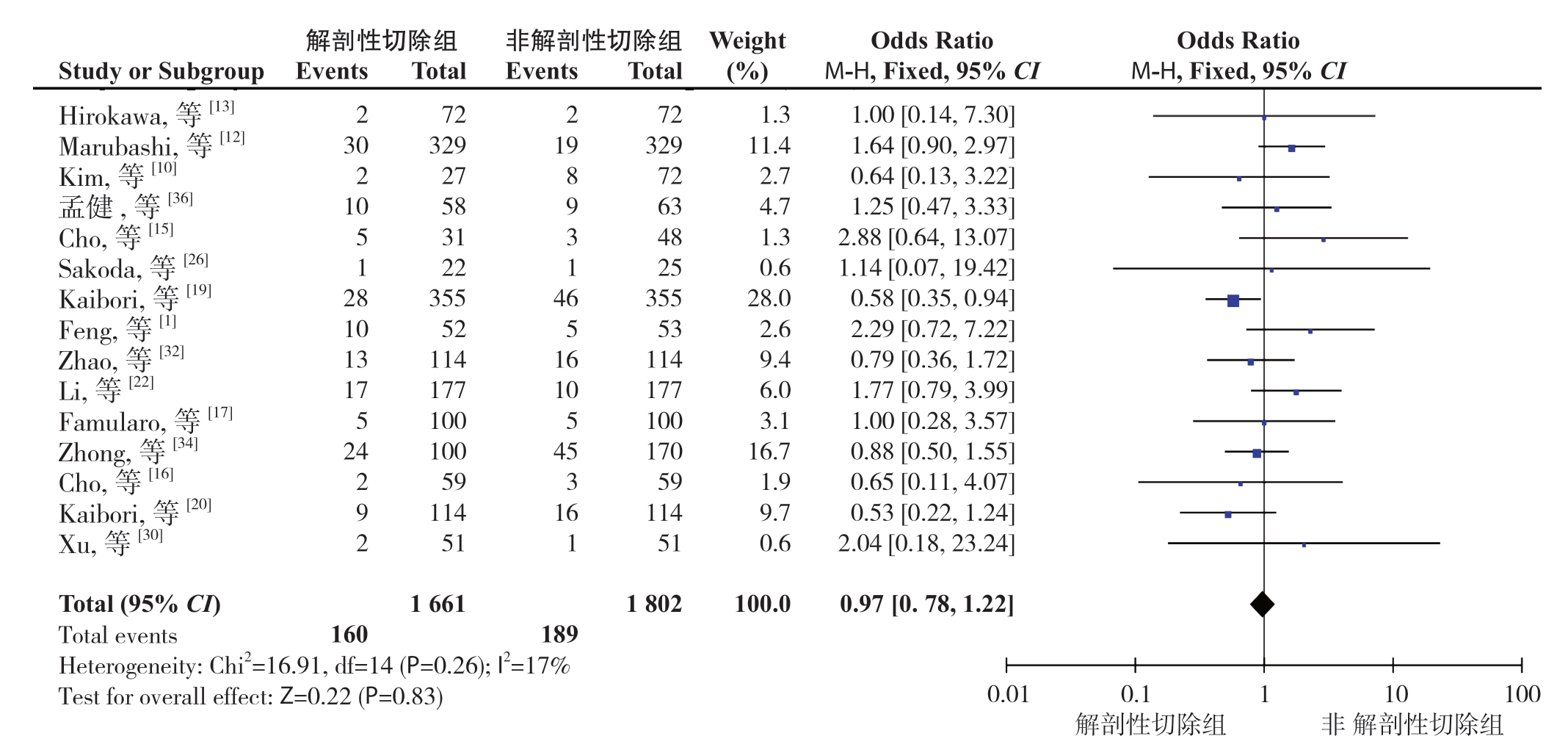
2.3.4 肝外转移发生率 共15 个研究报道了肝外转移发生率[1,10,12-13,15-17,19,20,22,26,30,32,34,36]。异质性检验显示各研究间无明显异质性(P=0.26,I2=17%),采用固定效应模型。Meta 分析结果显示:AR 与NAR 术后肝外转移发生率差异无统计学意义(OR=0.97,95% CI=0.78~1.22,P=0.83)(图5)。

图5 两组肝外转移发生率比较
Figure 5 Comparison of extrahepatic metastasis rates between the two groups
2.3.5 1、2、3、5年DFS 分别有32个研究[1,7,10-13,15-35,37-41]、29 个研究[1,7,10,12-13,15-24,27-35,37-41]、32 个研究[1,7,10-13,15-40]、26 个研究[1,7,10-13,15-21,23-29,31-33,37-38,40]报道了第1、2、3、5年DFS。异质性检验显示各研究间均无明显异质性(P=0.05,I2=31%;P=0.05,I2=32%;P=0.04,I2=33%;P=0.38,I2=6%),均采用固定效应模型。Meta 分析结果显示:与NAR 比较,AR 能明显提高患者术后1、2、3、5年DFS(OR=1.26,95% CI=1.12~1.42,P=0.0001;OR=1.31,95% CI= 1.17 ~1.46,P<0.00001;OR=1.32,95% CI= 1.19~1.48,P<0.00001;OR=1.40,95% CI= 1.23~1.58,P<0.00001)(图6)。
2.3.6 1、2、3、5年OS 分别有31个研究[1,7,10-13,15-16,18-35,37-41]、27 个研究[1,7,10,12-13,15-16,18-24,27-33,35,37-41]、31 个研究[1,7,10-13,15-16,18-40]、27 个研究[1,7,10-13,15-16,18-21,23-29,31-34,37-40]报道了第1、2、3、5年OS。异质性检验显示各研究间均无明显异质性(P=0.78,I2=0%;P=0.78,I2=0%;P=0.47,I2=0%;P=0.03,I2=37%),均采用固定效应模型。Meta 分析结果显示:AR 与NAR 术后1、2、3年OS 差异均无统计学意义(OR=1.01,95% CI=0.85 ~1.22,P=0.88;OR=1.15,95% CI=0.99 ~1.33,P=0.06;OR=1.13,95% CI=1.00~1.28,P=0.06)(图7A-C)。但与NAR 比较,AR 能提高患者术后5年OS(OR=1.16,95% CI=1.03~1.31,P=0.02)(图7D)。
2.3.7 手术时间等结局指标 除手术时间外其余次要结局指标差异均无统计学意义(表4)。
2.4 亚组分析结果
按照研究来源(国内、国外)和是否合并MVI进行亚组分析。亚组分析显示,1年DFS和2、3、5年OS结局指标在国内研究与国外研究中差异较大,具体表现为国内研究2年OS和3年OS在AR组中更高,国外研究1年DFS和5年OS两组中差异无统计学意义。再次复核数据提取无误后,分别做出漏斗图发现,国外研究5年OS和国内研究2年OS可能存在发表偏倚,其余结局指标未见明显发表偏倚。国内研究1年DFS和国外研究5年OS存在一定异质性,分别剔除宋泽兵等[37]和Kaibori等[20]研究异质性均明显降低但结果未发生方向性改变。此外,国内研究质量评价偏低,以6分为主,也可能是出现上述差异的原因之一,但目前尚无研究表明不同种族或地域会造成不同肝切除术后出现预后差异,有待进一步探索。固定效应模型Meta分析结果显示:合并MVI的研究间异质性降低,AR组在1、2、3、5年DFS及2、3、5年OS均高于NAR组,差异均有统计学意义(表5)。

图6 两组DFS 比较 A:1年DFS;B:2年DFS;C:3年DFS;D:5年DFS
Figure 6 Comparison of the DFS between the two groups A:1-year DFS;B:2-year DFS;C:3-year DFS;D:5-year DFS

图7 两组OS 比较 A:1年OS;B:2年OS;C:3年OS;D:5年OS
Figure 7 Comparison of the OS between the two groups A:1-year OS;B:2-year OS;C:3-year OS;D:5-year OS
表4 其他指标的Meta 分析结果
Table 4 Meta-analysis results of other variables

样本量(n)异质性检验结果结局指标纳入研究数效应模型Meta 分析结果试验组/对照组 I2(%)POR/MD95% CIP手术时间(min)10[1,12,16,19,22,23,34-36,41]1350/137593<0.00001 随机29.645.32~53.950.02术中出血量(mL)8[1,16,19,23,34,36,40,41]920/97779<0.0001随机32.83 -36.57~102.230.35输血率20[1,7,10,13,16,18-20,22-24,27-33,35,38]1934/1885310.09固定0.990.82~1.200.95术后并发症发生率15[10,17,19,22,23,27,29-31,33,35-38,41]1602/142200.45固定0.980.81~1.170.79住院时间(d)7[1,10,23,24,35,36,41]374/374610.02随机-0.28-1.82~1.250.72
表5 亚组分析
Table 5 Subgroup analysis

亚组纳入研究数异质性检验效应模型OR(95% CI)PI2(%)P 1年DFS国别国内14[1,11,22,28,30,32-35,37-41]390.07固定1.47(1.24~1.76)<0.0001国外18[7,10,12-13,15-21,23-27,29,31]50.40固定1.10(0.94~1.29)0.23是否合并MVI报告不明(混合)23[1,7,10-11,13,15-16,19-27,29-31,37-39,41]380.04固定1.28(1.09~1.49)0.002合并MVI9[12,17-18,28,32-35,40]200.27固定1.36(1.07~1.73)0.012年DFS国别国内13[1,22,28,30,32-35,37-41]290.16固定1.58(1.32~1.88)<0.00001国外16[7,10,12-13,15-21,23-24,27,29,31]130.31固定1.16(1.00~1.34)0.04是否合并MVI报告不明(混合)20[1,7,10,13,15-16,19-24,27,29-31,37-39,41]30.42固定1.32(1.14~1.53)0.0002合并MVI9[12,17-18,28,32-35,40]00.65固定1.55(1.23~1.95)0.00023年DFS国别国内14[1,11,22,28,30,32-40]460.03固定1.36(1.15~1.62)0.0004国外18[5,10,12-13,15-21,23-27,29,31]230.18固定1.30(1.12~1.49)0.0003是否合并MVI报告不明(混合)24[1,7,10-13,15-16,19-27,29-31,36-39]300.09固定1.29(1.14~1.47)0.0001合并MVI8[17-18,28,32-35,40]170.30固定1.78(1.38~2.30)<0.000015年DFS国别国内8[1,11,26,32-33,37-38,40]00.69固定1.61(1.29~2.01)<0.0001国外18[7,10,12-13,15-21,23-27,29,31]120.31固定1.31(1.12~1.52)0.0007是否合并MVI报告不明(混合)20[1,7,10-13,15-16,19-21,23-27,29,31,37-38]60.38固定1.29(1.11~1.49)0.0008合并MVI6[17-18,28,32-33,40]00.53固定2.07(1.46~2.94)<0.00011年OS国别国内14[1,11,22,28,30,32-35,37-41]00.82固定1.11(0.88~1.40)0.38国外17[7,10,12-13,15-16,18-21,23-27,29,31]00.59固定0.89(0.67~1.18)0.41是否合并MVI报告不明(混合)25[1,7,10-13,15-16,19-27,29-31,33,37-39,41]00.79固定0.95(0.76~1.18)0.62合并MVI6[18,28,32,34-35,40]00.50固定1.12(0.78~1.60)0.552年OS国别国内12[1,22,28,30,32-33,35,37-38,41]00.52固定1.25(1.01~1.54)0.04国外15[7,10,12-13,15-16,18-21,23-24,27,29,31]00.71固定1.06(0.86~1.30)0.58是否合并MVI报告不明(混合)22[1,7,10,12-13,15-16,19-24,27,29-31,33,37-39,41]00.66固定1.08(0.91~1.27)0.38合并MVI5[18,28,32,35,40]00.82固定1.54(1.07~2.21)0.02
表5 亚组分析(续)
Table 5 Subgroup analysis (continued)

亚组纳入研究数异质性检验效应模型OR(95% CI)PI2(%)P 3年OS国别国内14[1,11,22,28,30,32-40]20.42固定1.23(1.03~1.47)0.02国外17[7,10,12-13,15-16,18-21,23-27,29,31]00.52固定1.04(0.87~1.24)0.67是否合并MVI报告不明(混合)25[1,7,10-13,15-16,19-27,29-31,33,36-39]50.39固定1.05(0.91~1.22)0.52合并MVI6[18,28,32,34-35,40]00.58固定1.46(1.11~1.92)0.0075年OS国别国内10[1,11,28,32-34,37-40]130.32固定1.47(1.20~1.79)0.0002国外17[7,10,12-13,15-16,18-21,23-27,29,31]290.13固定1.01(0.86~1.17)0.94是否合并MVI报告不明(混合)22[1,7,10-13,15-16,19-21,23-27,29,31,33,37-39]420.02固定1.09(0.95~1.26)0.22合并MVI5[18,28,32,34,40]00.77固定1.52(1.13~2.03)0.005
2.5 敏感度分析结果
对异质性较大(I2>50%)的结局指标(早期复发率、肝内局部复发率、手术时间、术中出血量、住院时间)采用逐一剔除纳入研究的方法进行敏感度分析,以检验相关结果的稳定性。对于早期复发率和住院时间,逐一剔除研究后发现I2值变化不明显,提示结果较稳定;对于肝内局部复发率,剔除Shindoh等[27]研究后异质性明显降低(P=0.31,I2=16%),且研究结果未发生方向性改变(OR=0.45,95% CI=0.29~0.69,P=0.0002);对于手术时间,剔除孟健等[36]研究后异质性明显降低(P=0.0003,I2=72%),且研究结果未发生方向性改变(MD=20.95,95% CI=7.35 ~34.54,P=0.003);对于术中出血量,剔除胡文豪等[41]研究后异质性降低(P=0.02,I2=62%),且研究结果未发生方向性改变(MD=61.85,95% CI=-8.49~132.19,P=0.08)。
2.6 发表偏倚分析
发表偏倚通过漏斗图进行评估。选用纳入研究较多的3年OS这一结局指标为例,结果显示如下:各研究基本呈分布对称,提示无明显发表偏移(图8)。

图8 基于3年OS 的漏斗图
Figure 8 Funnel plot based on 3-year OS
3 讨论
HCC治疗性切除术始于1891年,经历了从“盲目”肝切除到“精准”肝切除的发展[5]。1954年法国医生Couinaud提出的肝脏八段法为AR奠定了解剖学基础。1985年Makuuchi等[44]提出并最早实践了AR对HCC的治疗,因此目前公认的AR概念也由此而来,即切除病灶并同时切除荷瘤肝段(亚肝段)及供应荷瘤肝段(亚肝段)的门静脉和肝动脉的主要分支。与之相对的NAR仅对切缘阴性有要求(一般≥1 cm),无关肝内解剖情况,能够保留较多的肝组织[5]。显然,AR在理论上强调完全切除肿瘤及荷瘤肝段,清除微小潜在病灶。
本研究表明,在手术彻底性方面,AR组术后肝内局部复发(同一肝段)率和早期复发(<2年)率较NAR组低,术后DFS和远期OS较NAR组高,表现出更好的治愈彻底性。HCC细胞易侵犯癌旁静脉系统,具有沿荷瘤肝段的门脉形成卫星灶进而早期复发和转移的生物学特性[45-47]。AR将荷瘤肝段及相应脉管系统一并切除,有效避免了沿门脉系统播散的卫星灶所致的早期局部扩散和转移,从而提高患者术后无瘤生存时间和远期总生存时间,减少了局部复发和早期复发[19,48]。Kaibori等[49]的研究也支持这一理论。但本研究也显示,在术后肝内远处复发(不同肝段)和肝外转移两术式无明显差异。其可能原因是HCC的起源是多中心的[50-51],即使切除了肝内宏观病灶,患者依然面临着远处转移和多中心复发的风险[1],而这种风险无法通过单纯的解剖性切除得以避免[52],同时通过局部扩散途径导致的远处复发在此时可以认为是微不足道的[53],因次AR的理论优势此时并未得到体现。本研究显示两种术式在近期(1、2、3年)OS中无明显差异。虽然AR减少了术后局部复发,但也降低了肝癌复发的抢救性治疗机会。同时本研究中肿瘤基本都处于早期,不同的术式对近期OS影响并不大,而AR对于OS的优势在远期才得以凸显[51,54]。不可忽略的一点是循环肿瘤细胞的全身迁移也是近期无瘤生存时间缩短和肿瘤肝外转移的原因[55],单纯解剖性切除尚不能阻止这一进程。上述结果,尤其近期与远期总生存率与之前Meta分析[56]的结果不一致,可能原因是其存在较大选择偏移[57],另外本研究纳入较多最新研究。对结论的解释尚需更多前瞻性的随机对照试验证实。
微血管是癌细胞克隆性增生首当其冲的侵袭部位,癌细胞形成的微癌栓增大、脱落并随血液全身迁移,是HCC患者术后复发和转移的重要原因[58]。MVI是影响HCC患者预后的独立危险因素[9,50,59]。有研究[60]报道合并MVI的HCC患者比非MVI患者中位生存期缩短30.4个月,累计生存率也差异明显。种种迹象表明,MVI是HCC细胞恶性程度增加、侵袭性增强的标志之一[58]。本研究显示,对合并MVI的HCC患者,AR较NAR在提高DFS和近远期OS均有明显优势。这与Famularo等[54]研究结论不同。基于将荷瘤肝段及其脉管系统全部切除的AR能最大限度地清除微小潜在病灶,降低患者肿瘤残留率和术后复发,因此在近期和远期生存率均显示出明显优势。该结论也很好地验证了AR的理论优势,并将这种优势转化为现实,应推荐合并MVI的HCC患者首选AR。值得注意的是,在上述一般HCC患者中,AR并没有在近期OS显示出优越性,这进一步从侧面凸显了AR对合并MVI的HCC患者的适用性和有效性。对合并MVI的HCC患者这一特殊人群的分析在一定程度上破解了之前多个研究在没有细化和分层的基础上探讨AR和NAR对患者DFS和OS的影响而结论不一的局面。但MVI是病理学诊断,术前不易获得,早期难以明确,限制了临床可操作性。近来,Lin等[61]提出了MVI术前预测模型,对预测MVI阳性的患者进行AR并取得一定成效。但至今尚未见独立预测指标的有关报道,这将是当下乃至未来一段时间研究的方向之一。
在手术时间和安全性方面,本研究显示,AR组的手术时间较NAR组长,其可能原因是:⑴AR操作难度大及术中辅助技术的引入需要花费较多时间;⑵不同中心对手术时间的定义未达成统一,可能造成两组手术时间差异;⑶分析表明,涉及手术时间的研究间异质性很大,因此手术时间的差异存在一定偏倚。但在术中出血量、输血率、术后肝衰竭等并发症发生率以及住院时间两术式均无统计学差异,在现有指标下表明AR安全可行。
本研究的局限性:⑴由于HCC合并MVI的原始研究较少,在亚组分析解释结果和推论时可能存在偏倚风险;⑵纳入研究中以小肝癌居多,涉及大肝癌的研究偏少,可能存在选择偏倚;⑶本研究中绝大部分患者肝功能Child-Pugh分级为A,另有少部分患者经过短期药物治疗后可从B恢复到A,因此结论限于肝功能良好的HCC患者;⑷纳入研究中仅有1篇RCT,结论尚需更多高质量前瞻性研究佐证。
综上所述,肝癌的手术切除是最大程度切除肿瘤侵犯区域和保留足够功能性肝组织间的精准平衡,即是彻底性和安全性的统一。在早期复发率居高不下的背景下,AR凸显出更好的治愈彻底性。尤其对合并MVI的HCC患者,AR术后近期和远期疗效均优于NAR,因此推荐在临床中实践。
[1]Feng X,Su Y,Zheng S,et al.A double blinded prospective randomized trial comparing the effect of anatomic versus nonanatomic resection on hepatocellular carcinoma recurrence[J].HPB(Oxford),2017,19(8):667-674.doi:10.1016/j.hpb.2017.04.010.
[2]刘勤,周少波.小肝癌射频消融与经腹腔镜切除治疗的近期疗效及成本效果分析[J].中国普通外科杂志,2020,29(5):596-602.doi:10.7659/j.issn.1005-6947.2020.05.011.
Liu Q,Zhou SB.Analysis of short-term efficacy and costeffectiveness of percutaneous radiofrequency ablation and laparoscopic hepatectomy in treatment of small hepatocellular carcinoma[J].Chinese Journal of General Surgery,2020,29(5):596-602.doi:10.7659/j.issn.1005-6947.2020.05.011.
[3]傅毅振,陈敏山.2019年原发性肝癌治疗进展[J].肿瘤综合治疗电子杂志,2020,6(2):86-89.doi:10.12151/JMCM.2020.02-11.
Fu YZ,Chen MS.Progress in the treatment of primary liver cancer in 2019[J].Journal of Multidisciplinary Cancer Management:Electronic Version,2020,6(2):86-89.doi:10.12151/JMCM.2020.02-11.
[4]吴周姜丽,李凯,张楷,等.单独或联合免疫疗法治疗肝癌的研究进展[J].中华肝脏病杂志,2020,28(6):471-474.doi:10.3760/cma.j.cn501113-20200520-00262.
Wu ZJL,Li K,Zhang K,et al.Research progress of immunotherapy alone and in combination for liver cancer[J].Chinese Journal of Hepatology,2020,28(6):471-474.doi:10.3760/cma.j.cn501113-20200520-00262.
[5]荚卫东,陈浩.肝癌肝切除术式选择的共识与争议[J].中国实用外科杂志,2018,38(4):353-356.doi:10.19538/j.cjps.issn1005-2208.2018.04.03.
Jia WD,Chen H.Controversy and consensus on the choice of hepatectomy for hepatocellular carcinoma[J].Chinese Journal of Practical Surgery,2018,38(4):353-356.doi:10.19538/j.cjps.issn1005-2208.2018.04.03.
[6]陈圣,赵继森,李靖华,等.肝细胞癌自噬相关长链非编码RNA预后模型的建立与分析[J].中国普通外科杂志,2020,29(7):839-848.doi:10.7659/j.issn.1005-6947.2020.07.008.Chen S,Zhao JS,Li JH,et al.Construction and analysis of prognostic model for hepatocellular carcinoma based on autophagy-related long non-coding RNAs[J].Chinese Journal of General Surgery,2020,29(7):839-848.doi:10.7659/j.issn.1005-6947.2020.07.008.
[7]Hokuto D,Nomi T,Yasuda S,et al.Does anatomic resection improve the postoperative outcomes of solitary hepatocellular carcinomas located on the liver surface?[J].Surgery,2018,163(2):285-290.doi:10.1016/j.surg.2017.08.024.
[8]Kang WH,Kim KH,Jung DH,et al.Long-term results of laparoscopic liver resection for the primary treatment of hepatocellular carcinoma:role of the surgeon in anatomical resection[J].Surg Endosc,2018,32(11):4481-4490.doi:10.1007/s00464-018-6194-0.
[9]Huang X,Lu S.A Meta-analysis comparing the effect of anatomical resection vs.non-anatomical resection on the long-term outcomes for patients undergoing hepatic resection for hepatocellular carcinoma[J].HPB (Oxford),2017,19(10):843-849.doi:10.1016/j.hpb.2017.06.003.
[10]Kim JM,Kwon CH,Joh JW,et al.Nonanatomical resection is comparable with anatomical resection in solitary hepatocellular carcinoma<5 cm in the right posterior section[J].Medicine (Baltimore),2016,95(51):e5382.doi:10.1097/MD.0000000000005382.
[11]林泽毅,朱少亮,黄山,等.单发小肝癌规则切除与非规则切除疗效及其预后因素分析[J].中华肿瘤防治杂志,2015,22(14):1128-1133.
Lin ZY,Zhu SL,Huang S,et al.Comparison of anatomic versus non-anatomic resection for small single hepatocellular carcinomas[J].Chinese Journal of Cancer Prevention and Treatment,2015,22(14):1128-1133.
[12]Marubashi S,Gotoh K,Akita H,et al.Anatomical versus nonanatomical resection for hepatocellular carcinoma[J].Br J Surg,2015,102(7):776-784.doi:10.1002/bjs.9815.
[13]Hirokawa F,Kubo S,Nagano H,et al.Do patients with small solitary hepatocellular carcinomas without macroscopically vascular invasion require anatomic resection? Propensity score analysis[J].Surgery,2015,157(1):27-36.doi:10.1016/j.surg.2014.06.080.
[14]赵首捷,杜锡林,杨振宇,等.微波消融辅助肝切除联合术后TACE治疗BCLC-B期肝细胞癌临床疗效分析[J].中国普通外科杂志,2020,29(7):812-819.doi:10.7659/j.issn.1005-6947.2020.07.005.
Zhao SJ,Du XL,Yang ZY,et al.Analysis of clinical efficacy of microwave ablation-assisted liver resection combined with postoperative TACE in treatment of BCLC-B stage hepatocellular carcinoma[J].Chinese Journal of General Surgery,2020,29(7):812-
819.doi:10.7659/j.issn.1005-6947.2020.07.005.
[15]Cho SH,Chun JM,Kwon HJ,et al.Outcomes and recurrence pattern after non-anatomic liver resection for solitary hepatocellular carcinomas[J].Korean J Hepatobiliary Pancreat Surg,2016,20(1):1-7.doi:10.14701/kjhbps.2016.20.1.1.
[16]Cho CW,Choi GS,Kim JM,et al.Long-Term Oncological Outcomes of Laparoscopic Liver Resection for Solitary Hepatocellular Carcinoma:Comparison of Anatomical and Nonanatomical Resection Using Propensity Score Matching Analysis[J].J Laparoendosc Adv Surg Tech A,2019,29(6):752-758.doi:10.1089/lap.2018.0600.
[17]Famularo S,Di Sandro S,Giani A,et al.Recurrence Patterns After Anatomic or Parenchyma-Sparing Liver Resection for Hepatocarcinoma in a Western Population of Cirrhotic Patients[J].Ann Surg Oncol,2018,25(13):3974-3981.doi:10.1245/s10434-018-6730-0.
[18]Hidaka M,Eguchi S,Okuda K,et al.Impact of Anatomical Resection for Hepatocellular Carcinoma With Microportal Invasion(vp1):A Multi-institutional Study by the Kyushu Study Group of Liver Surgery[J].Ann Surg,2020,271(2):339-346.doi:10.1097/SLA.0000000000002981.
[19]Kaibori M,Kon M,Kitawaki T,et al.Comparison of anatomic and non-anatomic hepatic resection for hepatocellular carcinoma[J].J Hepatobiliary Pancreat Sci,2017,24(11):616-626.doi:10.1002/jhbp.502.
[20]Kaibori M,Yoshii K,Hasegawa K,et al.Impact of systematic segmentectomy for small hepatocellular carcinoma[J].J Hepatobiliary Pancreat Sci,2020,27(6):331-341.doi:10.1002/jhbp.720.
[21]Kim S,Kim S,Song I,et al.Comparison of survival outcomes after anatomical resection and non-anatomical resection in patients with hepatocellular carcinoma[J].Korean J Hepatobiliary Pancreat Surg,2015,19(4):161-166.doi:10.14701/kjhbps.2015.19.4.161.
[22]Li SQ,Huang T,Shen SL,et al.Anatomical versus non-anatomical liver resection for hepatocellular carcinoma exceeding Milan criteria[J].Br J Surg,2017,104(1):118-127.doi:10.1002/bjs.10311.
[23]Matsumoto T,Kubota K,Aoki T,et al.Clinical Impact of Anatomical Liver Resection for Hepatocellular Carcinoma with Pathologically Proven Portal Vein Invasion[J].World J Surg,2016,40(2):402-411.doi:10.1007/s00268-015-3231-1.
[24]Molina-Romero FX,Palma-Zamora E,Morales-Soriano R,et al.Comparison of anatomical resection and non-anatomical resection in patients with hepatocellular carcinoma:propensity score matching method[J].Cir Cir,2019,87(3):328-336.doi:10.24875/CIRU.19000606.
[25]Okamura Y,Ito T,Sugiura T,et al.Anatomic versus nonanatomic hepatectomy for a solitary hepatocellular carcinoma :a casecontrolled study with propensity score matching[J].J Gastrointest Surg,2014,18(11):1994-2002.doi:10.1007/s11605-014-2646-6.
[26]Sakoda M,Ueno S,Iino S,et al.Survival Benefits of Small Anatomical Resection of the Liver for Patients with Hepatocellular Carcinoma and Impaired Liver Function,Based on New-Era Imaging Studies[J].J Cancer,2016,7(9):1029-1036.doi:10.7150/jca.15174.
[27]Shindoh J,Makuuchi M,Matsuyama Y,et al.Complete removal of the tumor-bearing portal territory decreases local tumor recurrence and improves disease-specific survival of patients with hepatocellular carcinoma[J].J Hepatol,2016,64(3):594-600.doi:10.1016/j.jhep.2015.10.015.
[28]Shi F,Zhou Z,Huang X,et al.Is anatomical resection necessary for early hepatocellular carcinoma? A single institution retrospective experience[J].Future Oncol,2019,15(17):2041-2051.doi:10.2217/fon-2019-0117.
[29]Viganò L,Procopio F,Mimmo A,et al.Oncologic superiority of anatomic resection of hepatocellular carcinoma by ultrasoundguided compression of the portal tributaries compared with nonanatomic resection:An analysis of patients matched for tumor characteristics and liver function[J].Surgery,2018,164(5):1006-1013.doi:10.1016/j.surg.2018.06.030.
[30]Xu H,Liu F,Hao X,et al.Laparoscopically anatomical versus non-anatomical liver resection for large hepatocellular carcinoma[J].HPB (Oxford),2020,22(1):136-143.doi:10.1016/j.hpb.2019.06.008.
[31]Yamamoto T,Yagi S,Uryuhara K,et al.Clinical factors that affect the outcomes after anatomical versus non-anatomical resection for hepatocellular carcinoma[J].Surg Today,2017,47(2):193-201.doi:10.1007/s00595-016-1397-2.
[32]Zhao H,Chen C,Gu S,et al.Anatomical versus non-anatomical resection for solitary hepatocellular carcinoma without macroscopic vascular invasion:A propensity score matching analysis[J].J Gastroenterol Hepatol,2017,32(4):870-878.doi:10.1111/jgh.13603.
[33]Zhao H,Ding WZ,Wang H,et al.Prognostic value of precise hepatic pedicle dissection in anatomical resection for patients with hepatocellular carcinoma[J].Medicine (Baltimore),2020,99(10):e19475.doi:10.1097/MD.0000000000019475.
[34]Zhong XP,Zhang YF,Mei J,et al.Anatomical versus Nonanatomical Resection for Hepatocellular Carcinoma with Microscope Vascular Invasion:A Propensity Score Matching Analysis[J].J Cancer,2019,10(17):3950-3957.doi:10.7150/jca.32592.
[35]刘煜,代扬,张欣雪,等.单发小肝癌合并微血管侵犯解剖性与非解剖性肝切除疗效对比分析[J].中华医学杂志,2018,98(24):1937-1940.doi:10.3760/cma.j.issn.0376-2491.2018.24.009.
Liu Y,Dai Y,Zhang XX,et al.Comparative analysis of anatomic and non-anatomic hepatectomy for single small hepatocellular carcinoma with microvascular invasion[J].National Medical Journal of China,2018,98(24):1937-1940.doi:10.3760/cma.j.issn.0376-2491.2018.24.009.
[36]孟健,杜松涛,张劲光.腹腔镜下肝楔形切除与解剖性切除的疗效对比研究[J].腹腔镜外科杂志,2016,21(5):321-324.doi:10.13499/j.cnki.fqjwkzz.2016.05.321.
Meng J,Du ST,Zhang JG.The comparison of curative effect between wedge and anatomical resection in laparoscopic liver resection[J].Journal of Laparoscopic Surgery,2016,21(5):321-324.doi:10.13499/j.cnki.fqjwkzz.2016.05.321.
[37]宋泽兵,胡继雄.肝脏第Ⅵ段肝硬化肝细胞癌患者行不同手术方式的预后比较[J].第三军医大学学报,2018,40(24):2266-2272.doi:10.16016/j.1000-5404.201807072.
Song ZB,Hu JX.Different surgical modalities for hepatocellular carcinoma confined to the Couinaud segment Ⅵ in cirrhotic patients:comparison of the outcomes[J].Journal of Third Military Medical University,2018,40(24):2266-2272.doi:10.16016/j.1000-5404.201807072.
[38]王宏伟,季顾惟,张慧,等.保留肝实质肝切除术治疗巴塞罗那A 期肝细胞癌的临床疗效及预后因素分析[J].中华消化外科杂志,2019,18(4):358-367.doi:10.3760/cma.j.issn.1673-9752.2019.04.011.
Wang HW,Ji GW,Zhang H,et al.Clinical efficacy of parenchymalsparing hepatectomy for barcelona clinic liver cancer stage A hepatocellular carcinoma and prognostic factors analysis[J].Chinese Journal of Digestive Surgery,2019,18(4):358-367.doi:10.3760/cma.j.issn.1673-9752.2019.04.011.
[39]罗翠松,林云,曾志峰,等.倾向值匹配法评价解剖性肝切除和非解剖性肝切除对肝细胞癌预后的影响[J].第二军医大学学报,2016,37(12):1568-1573.doi:10.16781/j.0258-879x.2016.12.1568.
Luo CS,Lin Y,Zeng ZF,et al.Propensity score-matching method for evaluating the effect of anatomic liver resection and nonanatomic liver resection on prognosis of hepatocellular carcinoma patients[J].Academic Journal of Second Military Medical University,2016,37(12):1568-1573.doi:10.16781/j.0258-879x.2016.12.1568.
[40]翟志超,任为正,刘志伟,等.解剖性与非解剖性肝切除治疗原发性肝癌远期疗效的比较[J].中华肝胆外科杂志,2017,23(11):771-775.doi:10.3760/cma.j.issn.1007-8118.2017.11.011.
Zhai ZC,Ren WZ,Liu ZW,et al.Prognosis of anatomic and non-anatomic liver resections for hepatocellular carcinoma[J].Chinese Journal of Hepatobiliary Surgery,2017,23(11):771-775.doi:10.3760/cma.j.issn.1007-8118.2017.11.011.
[41]胡文豪,王洪东,沈超楹,等.解剖性与非解剖性肝切除治疗原发性肝细胞癌临床疗效对比[J].中国现代手术学杂志,2019,23(3):172-178.doi:10.16260/j.cnki.1009-2188.2019.03.003.
Hu WH,Wang HD,Shen CY,et al.Comparison of Clinical Efficacy between Anatomical and Non-anatomical Hepatectomy for Primary Hepatocellular Carcinoma[J].Chinese Journal of Modern Operative Surgery,2019,23(3):172-178.doi:10.16260/j.cnki.1009-2188.2019.03.003.
[42]Stang A.Critical evaluation of the Newcastle-Ottawa scale for the assessment of the quality of nonrandomized studies in metaanalyses[J].Eur J Epidemiol,2010,25(9):603-605.doi:10.1007/s10654-010-9491-z.
[43]Higgins JPT,Green S.Cochrane handbook for systematic reviews of interventions version 5.1.0.The Cochrane Collaboration,2011[M].Available at:www.handbook.cochrane.org.
[44]Makuuchi M,Hasegawa H,Yamazaki S.Ultrasonically guided subsegmentectomy[J].Surg Gynecol Obstet,1985,161(4):346-350.
[45]Fan ST.Precise hepatectomy guided by the middle hepatic vein[J].Hepatobiliary Pancreat Dis Int,2007,6(4):430-434.
[46]Xu XF,Xing H,Han J,et al.Risk Factors,Patterns,and Outcomes of Late Recurrence After Liver Resection for Hepatocellular Carcinoma:A Multicenter Study From China[J].JAMA Surg,2019,154(3):209-217.doi:10.1001/jamasurg.2018.4334.
[47]Kim JM,Yi NJ,Kwon CHD,et al.Early disseminated recurrence after liver resection in solitary hepatocellular carcinoma[J].Ann Surg Treat Res,2018,94(3):129-134.doi:10.4174/astr.2018.94.3.129.
[48]解剖性与非解剖性肝切除术的选择[J].中国实用外科杂志,2018,38(4):418-419.doi:10.19538/j.cjps.issn1005-2208.2018.04.18.
Decision making between anatomic and non-anatomic liver resection[J].Chinese Journal of Practical Surgery,2018,38(4):418-419.doi:10.19538/j.cjps.issn1005-2208.2018.04.18.
[49]Kaibori M,Ishizaki M,Matsui K,et al.Predictors of microvascular invasion before hepatectomy for hepatocellular carcinoma[J].J Surg Oncol,2010,102(5):462-468.doi:10.1002/jso.21631.
[50]乐琪,朱同恩,莫志远,等.原发性肝癌患者手术切除术后早期复发影响因素分析[J].中国普通外科杂志,2019,28(1):18-23.doi:10.7659/j.issn.1005-6947.2019.01.003.
Le Q,Zhu TE,Mo ZY,et al.Analysis of factors for early recurrence of patients with hepatocellular carcinoma after resection[J].Chinese Journal of General Surgery,2019,28(1):18-23.doi:10.7659/j.issn.1005-6947.2019.01.003.
[51]Tan Y,Zhang W,Jiang L,et al.Efficacy and safety of anatomic resection versus nonanatomic resection in patients with hepatocellular carcinoma:A systemic review and meta-analysis[J].PLoS One,2017,12(10):e0186930.doi:10.1371/journal.pone.0186930.
[52]黄志勇,陈孝平.肝硬化与肝癌肝切除的科学决策[J].临床外科杂志,2019,27(8):631-633.doi:10.3969/j.issn.1005-6483.2019.08.001.
Huang ZY,Chen XP.Scientific decision-making in hepatectomy for liver cirrhosis and liver cancer[J].Journal of Clinical Surgery,2019,27(8):631-633.doi:10.3969/j.issn.1005-6483.2019.08.001.
[53]Marubashi S,Gotoh K,Akita H,et al.Analysis of Recurrence Patterns After Anatomical or Non-anatomical Resection for Hepatocellular Carcinoma[J].Ann Surg Oncol,2015,22(7):2243-2252.doi:10.1245/s10434-014-4214-4.
[54]Famularo S,Ceresoli M,Giani A,et al.Is It Just a Matter of Surgical Extension to Achieve the Cure of Hepatocarcinoma? A Meta-Analysis of Propensity-Matched and Randomized Studies for Anatomic Versus Parenchyma-Sparing Liver Resection[J].J Gastrointest Surg,2020.doi:10.1007/s11605-019-04494-5.[Online ahead of print]
[55]Rau KM,Liu CT,Hsiao YC,et al.Sequential Circulating Tumor Cell Counts in Patients with Locally Advanced or Metastatic Hepatocellular Carcinoma:Monitoring the Treatment Response[J].J Clin Med,2020,9(1):188.doi:10.3390/jcm9010188.
[56]Jiao S,Li G,Zhang D,et al.Anatomic versus non-anatomic resection for hepatocellular carcinoma,do we have an answer?A meta-analysis[J].Int J Surg,2020,80:243-255.doi:10.1016/j.ijsu.2020.05.008.
[57]Maduekwe UN,Charles A.An invited commentary on "Anatomic versus non-anatomic resection for hepatocellular carcinoma,do we have an answer? A meta-analysis"[Int.J.Surg.(2020) Epub ahead of print]:Does liver anatomy matter?[J].Int J Surg,2020,79:326.doi:10.1016/j.ijsu.2020.06.007.
[58]董辉,丛文铭.提高肝癌规范化病理诊断水平,为临床精细化治疗保驾护航-《原发性肝癌规范化病理诊断指南(2015年版)》解读[J].中国普通外科杂志,2016,25(7):939-943.doi:10.3978/j.issn.1005-6947.2016.07.001.
Dong H,Cong WM.Increasing the standardized pathology diagnosis level of primary hepatocellular carcinoma,escorting clinical precised treatment--Interpretation of "Guidelines for the diagnosis of primary hepatocellular carcinoma (2015)"[J].Chinese Journal of General Surgery,2016,25(7):939-943.doi:10.3978/j.issn.1005-6947.2016.07.001.
[59]杜培源,宋京海,乔江春,等.肝细胞癌患者微血管侵犯影响因素分析[J].中华肝胆外科杂志,2019,25(1):26-29.doi:10.3760/cma.j.issn.1007-8118.2019.01.007.
Du PY,Song JH,Qiao JC,et al.Influencing factors analysis of microvascular invasion in patients with hepatocellular carcinoma[J].Chinese Journal of Hepatobiliary Surgery,2019,25(1):26-29.doi:10.3760/cma.j.issn.1007-8118.2019.01.007.
[60]刘臻玉,武丹,区锦玲,等.肝癌微血管侵犯的特点及相关临床指标[J].肝胆胰外科杂志,2017,29(2):107-111.doi:10.11952/j.issn.1007-1954.2017.02.005.
Liu ZY,Wu D,Ou JL,et al.The characteristics and related biomarkers of microvascular invasion in hepatocellular carcinoma[J].Journal of Hepatopancreatobiliary Surgery,2017,29(2):107-111.doi:10.11952/j.issn.1007-1954.2017.02.005.
[61]Lin S,Ye F,Rong W,et al.Nomogram to Assist in Surgical Plan for Hepatocellular Carcinoma:a Prediction Model for Microvascular Invasion[J].J Gastrointest Surg,2019,23(12):2372-2382.doi:10.1007/s11605-019-04140-0.
