胆总管结石治疗困难的原因可大致分为患者因素和结石因素,患者因素包括一般情况差,高龄,出血倾向,消化道重建术后(内镜难以到达十二指肠乳头),乳头状憩室(胆管插管困难)、胆管狭窄等。结石因素包括巨大结石(>1.5 cm)、多发结石、形状异常、结石嵌顿和结石位置不佳等因素(表1)。为防止术中发生胆总管结石嵌顿并减少ERCP术后胰腺炎(post-ERCP pancreatitis,PEP)的发生,插管前必须先扩张胆管下端开口处的十二指肠乳头括约肌,主要方法有两种:经内镜下十二指肠乳头括约肌切开术(EST)和经内镜下球囊乳头扩张术(EPBD)[1-4]。EST最早于1974年开始实施[5],在内镜操作过程中摆正十二指肠乳头位置,注意切开方向、层次、深度以及合理使用点切方式可提高EST成功率,但EST可导致出血、穿孔以及十二指肠乳头狭窄、Oddi括约肌功能失调或丧失等并发症[1,6]。而EPBD使用直径6~10 mm球囊导管扩张Oddi括约肌,相对于EST,EPBD最大优势是无需切开Oddi括约肌从而可以保留十二指肠乳头的结构和功能完整,因而术后胆道感染和远期结石复发率更低[7-8],EPBD出血和穿孔风险也明显降低,即使是血液透析中、服用抗血栓药物或肝硬化等凝血功能异常者也可适用[9-10]。采用EPBD需根据结石最大径选择合适球囊导管扩张建立取石通道,因此EPBD也仅适用于球囊导管直径比扩张胆管直径更小的患者(表2)。然而,实际操作中由于各种原因仍存在内镜治疗困难的状况。
表1 胆总管结石治疗困难的原因
Table 1 Reasons for difficulties in choledocholithiasis treatment

患者因素结石因素健康状态解剖结构改变结石性状结石部位一般情况差消化道重建术后憩室内乳头巨大结石肝内胆管结石高龄乳头旁憩室多发小结石结石嵌顿出血倾向 胆管狭窄形状异常
表2 EPBD 球囊导管的选择方法
Table 2 Selection methods of EPBD balloon catheter
胆管直径6~8 mm8~10 mm 10~12 mm>12 mm球囊直径6 mm8 mm10 mmEPLBD
1 胆管插管困难
实施ERCP的前提是需成功进行胆管插管。虽然ERCP技术不断进步,但仍有3%~10%的患者胆管插管失败[11-12]。插管困难原因可大致归为两类:⑴术者因素(内镜技术难度高);⑵患者因素(到达十二指肠乳头的通路解剖结构异常、十二指肠乳头形态和位置改变、憩室内乳头、乳头开口狭窄、胆总管下端结石嵌顿等)。消化道重建术后和十二指肠乳头内结石嵌顿患者的十二指肠乳头位置改变因而内镜难以正面观察,如果内镜可正面观察到十二指肠乳头,乳头形态是否正常将成为影响插管成功与否的主要原因[13-14]。乳头形态可分为small、large、swollen(Hooknose)三类,后两类比第一类的插管难度约增加4.7倍[15]。因为胆管插管时间超过10 min插管次数>5次将导致PEP发病率明显升高[2,16],以往选择在胆管造影后行插管,10 min内插管率为53.9%~71.3%[17-18],成功率偏低。近年使用EST和用导丝引导插管法得以普及推广[19-20],在胰管中置入导丝引导插管(pancreatic guidewire cannulation,PGC)可协助固定并提起十二指肠乳头,使胆胰管更平直从而有利于显露操作视野。PGC分两类:单导丝法(single-guidewire technique,SGT)和双导丝法(double-guidewire technique,DGT)。SGT插管困难时,可先用极少量造影剂进行胰头部胰管造影,确认主胰管至分支开口处,向主胰管内插入导丝,插管时如导丝有抵触感或者导丝前端弯曲,而胰管内无梗阻时,则导丝可能已进入主胰管分支,此时可反复进行先后退再微调整动作插入导丝,必要时可追加少量造影剂确认主胰管走形,避免插入主胰管分支[21]。与胆管造影法相比,PGC法的胆管插管成功率更高,而PEP发病率更低[22-23]。但对于插管困难患者,如果固执地仅使用PGC法反复插管也可导致PEP发病率增加[22],因此,在实际操作时也可考虑联用胆管造影法提高插管成功率。另一方面,EST法单次胆管插管成功率约86.7%,熟练操作者PEP发病率低至2.6%[24]。有RCT显示PGC和EST两者在插管成功率方面无差异,但PGC所诱发的PEP发病率较EST高[25]。综上所述,对于胆管插管困难患者,如果术前预计将使用EST,术中首选EST,注意EST联用EPBD可提高插管成功率,如存在乳头解剖结构异常、门脉高压、凝血功能异常等困难,则单用EPBD或者PGC法插管,但应尽量避免长时间反复行胆管插管。
以上方法均失败时,可采用超声内镜引导胆道会师术(EUS-RV)引导胆管插管,EUS-RV是指在超声内镜(EUS)引导下经胃或十二指肠穿刺胆管,导丝插入胆管后经乳头进入十二指肠,从而引导内镜治疗的技术[26]。EUS-RV胆管插管成功率58%~98%,联用EPBD时插管成功率更高[27]。但对于消化道重建术后(毕II式、Roux-en-Y胃空肠吻合术)患者,因常规小肠镜很难到达十二指肠乳头,EUS-RV法应用困难[28]。另外,EUS-RV技术难度高,需麻醉配合,插管成功率较EST/EPBD更低[19],目前EUS-RV多适用于ERCP插管失败患者。
2 高龄/一般情况差者
对于合并多种内科疾病的年老体弱胆总管结石患者,长时间ERCP操作耐受性差,手术治疗风险高病死率高,内镜治疗力求快速简便且遵循最小“损伤控制”原则[29-30]。比如,出血是EST的常见并发症,而EPBD出血风险更低可安全应用于高龄患者[9-10],但高龄患者乳头萎缩使EST和EPBD难度加大[31]。临床上对于高龄且一般情况差者,术前评估患者无法耐受取石或结石多发或直径较大时,多采用内镜下胆道引流术(endoscopic retrograde biliary drainage,ERBD),置入支架或鼻胆管保持胆汁引流通畅[4,32],ERBD具备无需全身麻醉、手术时间短、恢复快等优点,放置鼻胆管引流待一般情况改善后择期取石[33]。而胆管内留置支架并配合口服药物溶石或者置入载药胆管支架溶石治疗难治性胆总管结石也备受期待[34]。对于常规ERCP置入胆管支架困难患者,可采用EUSRV技术穿刺胆管并留置支架。但Bergman等[35]对58例实施ERBD术后老年患者进行随访,40%患者于术后36个月左右出现了以胆管炎为主的远期并发症,死亡44例中的9例与胆管炎有关。另有研究发现,长期放置塑料支架可导致支架移位堵塞胆道[36],且可导致支架-结石复合体(stent-stone complex,SSC)形成,诱发胆管炎并加速胆管结石形成[37]。因此仅留置胆管支架无法解决根本问题,去除结石仍是基本治疗原则[38]。
3 消化道重建术后
对于消化道重建术后(Billroth I式、Billroth II式、或胃空肠Roux-en-Y吻合、胆管空肠Roux-en-Y吻合)患者行ERCP时,在内镜到达十二指肠乳头和对胆管插管入路判断这两点上较困难[13,39]。Billroth I式吻合术后消化道长度缩短,内镜容易进入十二指肠,ERCP流程基本相同。Billroth II式吻合方式可分为结肠前和结肠后,结肠后途径由于输入襻短,小肠镜可到达十二指肠乳头,但由于结肠前途径输入襻长,通常使用的小肠镜则很难到达十二指肠乳头[40]。胃空肠Roux-en-Y吻合和胆管空肠Roux-en-Y吻合术后到达十二指肠乳头和胆肠吻合口的插管距离过长,普通内镜无法到达[41]。以往对于此类患者只能选择经皮经肝胆道镜(PTCS)或外科手术。其中,PTCS下EHL/激光碎石比外科手术创伤更小,可作为第一代替手段,但也存在治疗周期长,结石取净率低等缺点[42]。普通小肠镜因为视野窄、硬度高、直径大、长度不够等原因,应用时难以跨越吻合口到达乳头或胆肠吻合口,近年使用镜身更长的单气囊(single-balloon enteroscopy,SBE)或双气囊小肠镜(double-balloon enteroscopy,DBE)行ERCP到达十二指肠乳头和胆管空肠吻合口成功率明显提高,Billroth II式胃切除术患者成功率50%~92%,Roux-en-Y吻合患者成功率在33%~67%之间[43-44]。而对于SBE/DBE无法到达乳头者,可采用EUS-AG和PTCS下EHL/激光碎石取石[45-46]。另外,即使内镜到达十二指肠乳头,由于乳头上下位置颠倒呈倒乳头型,胆管解剖位置反向,加之插管方向与常规ERCP方向不同甚至相反,造影导管难以对准乳头开口,插管难度高[2]。而EST应用于消化道重建术后患者操作难度大风险高,EPBD和PGC则相对易操作且安全[10]。近期,在EUS引导下,从残胃或空肠上段导丝穿刺入左肝内胆管,再沿胆管进入胆总管,到达十二指肠乳头,用球囊扩张十二指肠乳头后,将结石推入十二指肠腔的EUS引导下顺行性治疗(EUS-guided antegrade technique,EUS-AG)成功案列也逐渐增加,但需注意消化道穿孔、胆汁漏等的发生[47]。
4 巨大结石
通常,如果结石直径<1 cm,根据EST及其后使用的取石网篮或球囊导管的标准ERCP术能达到治疗目的,对于直径1.5~2.0 cm的巨大结石,直接使用取石网篮套取非常困难[4]。内镜下机械碎石(endoscopic mechanical lithotripsy,EML)由于简便、廉价、碎石率高(84%~98%),以往作为碎石首选[48]。而对于直径>2cm巨大结石或填充型结石,EML碎石后取石难度高、易复发[49]。体外冲击波碎石疗法(extracorporeal shock wave lithotripsy,ESWL)和激光/液电碎石(electrohydraulic lithotripsy,EHL)适用于胆总管巨大结石时碎石率在73%~96%之间,碎石后可通过自然排石(<10%)或内镜取石[50-52]。但ESWL碎石组织损伤大,术前经ENBD管行胆道造影可准确定位结石以减少碎石时引起的组织损伤,也可经ENBD管应用肾上腺素和止血药物冲洗防治ESWL术后胆道出血[53]。尽管如此,ESWL总的并发症发生率仍偏高,目前已较少应用于临床。经口胆道镜(peroral cholangioscopy,POCS)下EHL碎石率为85%~98%,结石取净率为64%~97%,整体疗效优于ESWL[46],这类POCS技术采用子母胆道镜方式,通过母镜钳孔,向胆管内插入细径胆道内镜,再从钳孔中插入激光或EHL探针碎石,但此子母镜系统需双人操作,仅可两方向转位,且子镜昂贵易受损。近年Boston Scientific公司新开发了一种手术视野清晰、四方向转位、有独立冲洗通道的单人操作SpyGlass胆道镜系统,该系统是在胆道子母镜基础上开发的胰胆管疾病诊疗系统,可对肝内外胆管清晰观察和取材,SpyGlass胆道镜系统直视下激光碎石/EHL可用于常规内镜无法碎石取石的患者,结石取净率在73%~91%之间,进镜前行EST或EPBD可提高SpyGlass传送导管进入胆道成功率,进镜后冲洗胆道时不宜压力过大,以免小结石进入肝内胆管造成残留,虽然此系统对于使用常规胆道镜无法完成的胆总管巨大结石患者安全有效,但SpyGlass胆道镜系统存在极易受损、技术难度高且费用高昂等局限性,临床普及困难[54]。另一种非子母胆道镜方式,使用超细径鼻内镜直接插入胆管内的Direct POCS的结石取净率在85%~89%之间,但该方法操作复杂,不适用于消化道重建术后患者[55]。
对于大结石,理论上取石之前十二指肠乳头扩张越大,插入碎石取石设备的难度就越低。内镜下大口径球囊乳头扩张术(endoscopic papillary large balloon dilation,EPLBD)使用比EPBD标准使用的直径6~10 mm更大的直径为12~20 mm球囊导管扩张十二指肠乳头,多用于清除直径>12 mm的胆总管单发结石或多发结石[56],EPLBD联用EST结石取净率在94%~100%之间,而并发症发生率无明显升高[57]。而且,因为EPLBD容易去除乳头扩张后的结石,提高了单次结石取净率并降低了手术时间长所导致的高并发症发生率[58]。而EPLBD并发症如出血和穿孔发生率仅0~9%,PEP发生率与EST相比无明显升高[59]。但EST行较大范围切开乳头括约肌的术式相较于EPLBD的并发症发生率却明显偏高,且EST术后远期胆管炎发生率和胆管结石复发率更高[49]。EPLBD对于胆管内大结石、多发结石简单有效,临床如无EST禁忌,多采用先EST小范围切开后EPLBD的方法,单次结石取净率更高且可减少单用EPLBD所造成的胰管受压诱发PEP发生[60]。因此,对于胆总管巨大结石和多发结石,EST+EPLBD 法常作为一线治疗方案。
5 结石嵌顿
胆总管结石嵌顿可导致胆道完全梗阻,诱发急性化脓性胆管炎、感染性休克等严重并发症。胆总管末端结石嵌顿引起胆道轴线异常、乳头开口移位、成角,导致导丝、球囊导管和网篮无法越过结石进入远端胆管,常规ERCP和EST插管难度大,治疗方法应根据结石性状、局部解剖以及患者全身情况等进行选择,避免长时间反复插管或取石诱发胆胰汇合部损伤、出血和穿孔等。结石嵌顿时由于结石卡压和结石上方胆管明显扩张,乳头开窗术有助于提高插管成功率,乳头开窗后采用合适的取石方式更为重要[61]。Tsuyuguchi等[62]对122例治疗困难的胆管结石(Mirizzi征候群McSherry I型3例,II型50例,嵌顿结石50例,巨大结石19例)行POCS下EHL/激光碎石,Mirizzi征候群II型嵌顿结石和巨大结石取净率96%,但结石未在胆总管中的Mirizzi征候群I型治疗均告失败。如胆总管末端结石嵌顿合并胆总管末端狭窄,应根据狭窄长度选择EST 或Oddi括约肌切开成形术,但须注意Oddi括约肌切开成形术可破坏括约肌功能。如嵌顿结石体积大且质地坚硬,碎石不彻底导致网篮套取结石后无法脱出时,应反复活动网篮,避免盲目向外牵拉导致胆道出血或穿孔,必要时转开腹手术。
6 肝内胆管结石
肝内胆管结石常伴有胆管狭窄、结石嵌顿,或由于胆管屈曲,内镜取石非常困难。加之结石复发率和胆管癌的合并率高,所以外科手术切除病变区域的部分肝脏为治疗原则[63]。但是,对于一般情况不佳、拒绝手术、大范围多发结石等原因而无法耐受肝切除手术的患者,可行PTCS下EHL/激光碎石,并发症包括肝胆管裂伤、腹腔脓肿、胆道出血、感染性休克等,其结石以及胆管炎复发率约35%~63%[64]。但是行PTCS下EHL/激光碎石术需在经皮经肝窦道形成之后才能进行,窦道形成时间长达2周以上,治疗期间存在长期留置经皮经肝引流管导致患者腹痛、发热以及生活不便等缺点。近年来,SpyGlass引导下激光碎石疗法正逐渐应用于肝内胆管一级分支结石的治疗,但因肝内胆管结石的多发性以及常伴有胆管迂曲狭窄等原因,其应用范围以及治疗效果有限[65]。Iwashita等[28]报道了应用EUS-RV经胆管穿刺进入肝内引导EHL碎石的术式可用于肝内胆管结石的治疗,但操作难度极高,且对于肝内胆管多发结石患者结石取净非常困难,此方法的推广应用尚待进一步研究改进。
7 小结
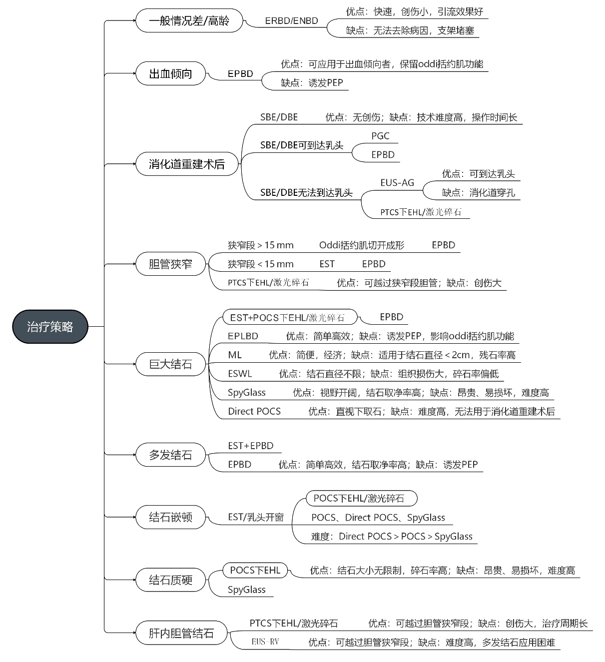
笔者将困难型胆总管结石的内镜治疗方案及优缺点列举于图1。对于全身状态不佳或危重患者,首选ERBD或ENBD解除梗阻,不强调一次性取石。凝血功能不佳者采用EPBD。对于内镜较难达到十二指肠乳头和胆肠吻合口的消化道重建患者,选择SBE/DBE或者EUS-AG,如两者均失败,行PTCS下EHL/激光碎石。胆管狭窄患者根据狭窄段长度分别采用Oddi括约肌切开成形术或者EST,之后采用EPBD插管取石,如失败,单独采用PTCS下EHL/激光碎石。ML、ESWL、SpyGlass和Direc POCS均可用于巨大结石碎石取石,但可优先采用先EST后POCS下EHL/激光碎石术,联用EPLBD可提高结石取净率。对于多发结石,采用先EST后EPBD,如结石多发但体积小,可单用EPBD。对于胆胰汇合部胆石和Mirizzi综合征(McSherry II型),选择先EST或乳头开窗术,再优先采用POCS下EHL/激光碎石。对肝内胆管结石,PTCS下EHL/激光碎石仍是最主要选项,EUS-RV可做备用选项。

图1 难型胆总管结石的内镜治疗方案及优缺点
Figure 1 Endoscopic treatment strategies for the complex choledocholithiasis and their advantages and disadvantages
文中总结了困难型胆总管结石的内镜治疗各种策略以及优缺点,将有助于临床工作中正确选择难治型胆总管结石的最佳内镜方案,解决病因并尽力减轻患者痛苦以及经济负担。
[1]Ryozawa S,Itoi T,Katanuma A,et al.Japan Gastroenterological Endoscopy Society guidelines for endoscopic sphincterotomy[J].Dig Endosc,2018,30(2):149-173.doi:10.1111/den.13001.
[2]Mine T,Morizane T,Kawaguchi Y,et al.Clinical practice guideline for post-ERCP pancreatitis[J].J Gastroenterol,2017,52(9):1013-1022.doi:10.1007/s00535-017-1359-5.
[3]Fujisawa T,Kagawa K,Hisatomi K,et al.Is endoscopic papillary balloon dilatation really a risk factor for post-ERCP pancreatitis?[J].World J Gastroenterol,2016,22(26):5909-5916.doi:10.3748/wjg.v22.i26.5909.
[4]杨文,李修红.ERCP联合EST治疗急性胆源性胰腺炎的临床效果与适应证分析[J].中国普通外科杂志,2020,29(9):1119-1125.doi:10.7659/j.issn.1005-6947.2020.09.013.
Yang W,Li XH.Analysis of clinical efficacy and indications of ERCP combined with EST for treatment of acute biliary pancreatitis[J].Chinese Journal of General Surgery,2020,29(9):1119-1125.doi:10.7659/j.issn.1005-6947.2020.09.013.
[5]Kawai K,Akasaka Y,Murakami K,et al.Endoscopic sphincterotomy of the ampulla of Vater[J].Gastrointest Endosc,1974,20(4):148-151.doi:10.1016/s0016-5107(74)73914-1.
[6]Köksal AŞ,Eminler AT,Parlak E.Biliary endoscopic sphincterotomy:Techniques and complications[J].World J Clin Cases,2018,6(16):1073-1086.doi:10.12998/wjcc.v6.i16.1073.
[7]Cheon YK,Lee TY,Kim SN,et al.Impact of endoscopic papillary large-balloon dilation on sphincter of Oddi function:a prospective randomized study[J].Gastrointest Endosc,2017,85(4):782-790.doi:10.1016/j.gie.2016.08.031.
[8]叶诚,张辉,周文策.内镜逆行胰胆管造影术后胆总管结石复发的相关因素研究进展[J].中国普通外科杂志,2019,28(8):995-999.doi:10.7659/j.issn.1005-6947.2019.08.013.
Ye C,Zhang H,Zhou WC.Factors for recurrence of choledocholithiasis after endoscopic retrograde cholangiopancreatography:recent progress[J].Chinese Journal of General Surgery,2019,28(8):995-999.doi:10.7659/j.issn.1005-6947.2019.08.013.
[9]Hung TH,Tseng CW,Chen YC et al.Endoscopic papillary balloon dilation decreases the risk of bleeding in cirrhotic patients compared with endoscopic biliary sphincterotomy:A national populationbased study[J].Medicine (Baltimore),2019,98(30):e16529.doi:10.1097/MD.0000000000016529.
[10]Kuo CM,Chiu YC,Liang CM,et al.The efficacy of limited endoscopic sphincterotomy plus endoscopic papillary large balloon dilation for removal of large bile duct stones[J].BMC Gastroenterol,2019,19(1):93.doi:10.1186/s12876-019-1017-x.
[11]Cunningham JT.The Art of Selective Cannulation at ERCP[J].Curr Gastroenterol Rep,2019,21(2):7.doi:10.1007/s11894-019-0673-x.
[12]Berry R,Han JY,Tabibian JH.Difficult biliary cannulation:Historical perspective,practical updates,and guide for the endoscopist[J].World J Gastrointest Endosc,2019,11(1):5-21.doi:10.4253/wjge.v11.i1.5.
[13]Park TY,Bang CS,Choi SH,et al.Forward-viewing endoscope for ERCP in patients with Billroth II gastrectomy:a systematic review and meta-analysis[J].Surg Endosc,2018,32(11):4598-4613.doi:10.1007/s00464-018-6213-1.
[14]Park TY,Song TJ.Recent advances in endoscopic retrograde cholangiopancreatography in Billroth II gastrectomy patients:A systematic review[J].World J Gastroenterol,2019,25(24):3091-3107.doi:10.3748/wjg.v25.i24.3091.
[15]Haraldsson E,Kylänpää L,Grönroos J,et al.Macroscopic appearance of the major duodenal papilla influences bile duct cannulation:a prospective multicenter study by the Scandinavian Association for Digestive Endoscopy Study Group for ERCP[J].Gastrointest Endosc,2019,90(6):957-963.doi:10.1016/j.gie.2019.07.014.
[16]Tryliskyy Y,Bryce GJ.Post-ERCP pancreatitis:Pathophysiology,early identification and risk stratification[J].Adv Clin Exp Med,2018,27(1):149-154.doi:10.17219/acem/66773.
[17]Katsinelos P,Paroutoglou G,Kountouras J,et al.A comparative study of standard ERCP catheter and hydrophilic guide wire in the selective cannulation of the common bile duct[J].Endoscopy,2008,40(4):302-307.doi:10.1055/s-2007-995483.
[18]Ishii S,Fujisawa T,Isayama H,et al.Clinical Evaluation of a Newly Developed Guidewire for Pancreatobiliary Endoscopy[J].J Clin Med,2020,9(12):4059.doi:10.3390/jcm9124059.
[19]Testoni PA,Mariani A,Aabakken L,et al.Papillary cannulation and sphincterotomy techniques at ERCP:European Society of Gastrointestinal Endoscopy (ESGE) Clinical Guideline[J].Endoscopy,2016,48(7):657-683.doi:10.1055/s-0042-108641.
[20]Sugimoto M,Takagi T,Suzuki R,et al.Pancreatic stents for the prevention of post-endoscopic retrograde cholangiopancreatography pancreatitis should be inserted up to the pancreatic body or tail[J].World J Gastroenterol,2018,24(22):2392-2399.doi:10.3748/wjg.v24.i22.2392.
[21]Franzini TP,Rocha RSP,Guedes HG,et al.Single-guidewire double-tip cannulation for difficult biliary access:the DTC technique[J].Endoscopy,2018,50(8):E212-213.doi:10.1055/a-0624-1280.
[22]Tse F,Yuan Y,Moayyedi P,et al.Double-guidewire technique in difficult biliary cannulation for the prevention of post-ERCP pancreatitis:a systematic review and meta-analysis[J].Endoscopy,2017,49(1):15-26.doi:10.1055/s-0042-119035.
[23]Kwon CI,Koh DH,Song TJ,et al.Technical Reports of Endoscopic Retrograde Cholangiopancreatography Guidewires on the Basis of Physical Properties[J].Clin Endosc,2020,53(1):65-72.doi:10.5946/ce.2019.114.
[24]Sundaralingam P,Masson P,Bourke MJ.Early Precut Sphincterotomy Does Not Increase Risk During Endoscopic Retrograde Cholangiopancreatography in Patients With Difficult Biliary Access:A Meta-analysis of Randomized Controlled Trials[J].Clin Gastroenterol Hepatol,2015,13(10):1722-1729.doi:10.1016/j.cgh.2015.06.035.
[25]Huang L,Yu QS,Zhang Q,et al.Comparison between doubleguidewire technique and transpancreatic sphincterotomy technique for difficult biliary cannulation[J].Dig Endosc,2015,27(3):381-387.doi:10.1111/den.12387.
[26]Itoi T,Dhir V.EUS-guided biliary rendezvous:slow,hesitant,baby steps forward[J].Gastrointest Endosc,2016,83(2):401-403.doi:10.1016/j.gie.2015.05.049.
[27]Matsubara S,Nakagawa K,Suda K,et al.A Proposed Algorithm for Endoscopic Ultrasound-Guided Rendezvous Technique in Failed Biliary Cannulation[J].J Clin Med,2020,9(12):3879.doi:10.3390/jcm9123879.
[28]Iwashita T,Uemura S,Yoshida K,et al.EUS-guided hybrid rendezvous technique as salvage for standard rendezvous with intrahepatic bile duct approach[J].PLoS One,2018,13(8):e0202445.doi:10.1371/journal.pone.0202445.
[29]李婧伊,刘飞,马跃峰,等.年龄对ERCP治疗胆总 管结石术后并发胰腺炎及严重程度的影响[J].中国普通外科杂志,2019,28(8):936-942.doi:10.7659/j.issn.1005-6947.2019.08.005.
Li JY,Liu F,Ma YF,et al.Influence of age on postoperative pancreatitis and its severity after ERCP for choledocholithiasis[J].Chinese Journal of General Surgery,2019,28(8):936-942.doi:10.7659/j.issn.1005-6947.2019.08.005.
[30]Nassar Y,Richter S.Management of complicated gallstones in the elderly:comparing surgical and non-surgical treatment options[J].Gastroenterol Rep (Oxf),2019,7(3):205-211.doi:10.1093/gastro/goy046.
[31]Saito H,Koga T,Sakaguchi M,et al.Safety and Efficacy of Endoscopic Removal of Common Bile Duct Stones in Elderly Patients ≥90 Years of Age[J].Intern Med,2019,58(15):2125-2132.doi:10.2169/internalmedicine.2546-18.
[32]杨勇,田明国,辛国军,等.C/S-J型胆道自行脱落支架在经内镜逆行胰胆管造影术后胆道引流中应用[J].中国普通外科杂志,2019,28(8):929-935.doi:10.7659/j.issn.1005-6947.2019.08.004.
Yang Y,Tian MG,Xin GJ,et al.Application of C/S-J type selfreleasing biliary stent for biliary drainage after endoscopic retrograde cholangiopancreatography[J].Chinese Journal of General Surgery,2019,28(8):929-935.doi:10.7659/j.is sn.1005-6947.2019.08.004.
[33]Yin P,Wang M,Qin R,et al.Intraoperative endoscopic nasobiliary drainage over primary closure of the common bile duct for choledocholithiasis combined with cholecystolithiasis:a cohort study of 211 cases[J].Surg Endosc,2017,31(8):3219-3226.doi:10.1007/s00464-016-5348-1.
[34]Huang C,Cai XB,Guo LL,et al.Drug-eluting fully covered selfexpanding metal stent for dissolution of bile duct stones in vitro[J].World J Gastroenterol,2019,25(26):3370-3379.doi:10.3748/wjg.v25.i26.3370.
[35]Bergman JJ,Rauws EA,Tijssen JG,et al.Biliary endoprostheses in elderly patients with endoscopically irretrievable common bile duct stones:report on 117 patients[J].Gastrointest Endosc,1995,42(3):195-201.doi:10.1016/s0016-5107(95)70091-9.
[36]Taj MA,Ghazanfar S,Qureshi S,et al.Plastic stent migration in ERCP;a tertiary care experience[J].J Pak Med Assoc,2019,69(8):1099-1102.
[37]Kaneko J,Kawata K,Watanabe S,et al.Clinical characteristics and risk factors for stent-stone complex formation following biliary plastic stent placement in patients with common bile duct stones[J].J Hepatobiliary Pancreat Sci,2018,25(10):448-454.doi:10.1002/jhbp.584.
[38]Iida T,Kaneto H,Wagatsuma K,et al.Efficacy and safety of endoscopic procedures for common bile duct stones in patients aged 85 years or older:A retrospective study[J].PLoS One,2018,13(1):e0190665.doi:10.1371/journal.pone.0190665.
[39]Aiolfi A,Asti E,Rausa E,et al.Trans-Gastric ERCP After Rouxen-Y Gastric Bypass:Systematic Review and Meta-Analysis[J].Obes Surg,2018,28(9):2836-2843.doi:10.1007/s11695-018-3258-0.
[40]El Hajj II,Al-Haddad M.Challenges in ERCP post-Billroth II gastrectomy:Is it the scope,tools or technique?[J]Saudi J Gastroenterol,2019,25(6):333-334.doi:10.4103/sjg.SJG_583_19.
[41]Ali MF,Modayil R,Gurram KC,et al.Spiral enteroscopy-assisted ERCP in bariatric-length Roux-en-Y anatomy:a large single-center series and review of the literature[J].Gastrointest Endosc,2018,87(5):1241-1247.doi:10.1016/j.gie.2017.12.024.
[42]Yamamoto R,Abe T,Hosaka S,et al.Intrahepatic bile duct stone treatment combining electrohydraulic lithotripsy and peroral cholangioscopy through a choledochoduodenal fistula[J].Endoscopy,2019,51(10):E290-292.doi:10.1055/a-0914-2581.
[43]Sano I,Katanuma A,Kuwatani M,et al.Long-term outcomes after therapeutic endoscopic retrograde cholangiopancreatography using balloon-assisted enteroscopy for anastomotic stenosis of choledochojejunostomy/pancreaticojejunostomy[J].J Gastroenterol Hepatol,2019,34(3):612-619.doi:10.1111/jgh.14605.
[44]Hashimoto R,Matsuda T,Aoki H,et al.Double-balloon assisted trans-anal ERCP in a patient with Roux-en-Y choledochojejunostomy[J].Gastrointest Endosc,2017,85(6):1298-1299.doi:10.1016/j.gie.2016.07.059.
[45]Mora Soler AM,Álvarez Delgado A,Piñero Pérez MC,et al.Endoscopic ultrasound-guided choledochoduodenostomy after a failed or impossible ERCP[J].Rev Esp Enferm Dig,2018,110(5):299-305.doi:10.17235/reed.2018.5040/2017.
[46]Kamiyama R,Ogura T,Okuda A,et al.Electrohydraulic Lithotripsy for Difficult Bile Duct Stones under Endoscopic Retrograde Cholangiopancreatography and Peroral Transluminal Cholangioscopy Guidance[J].Gut Liver,2018,12(4):457-462.doi:10.5009/gnl17352.
[47]Bukhari M,Kowalski T,Nieto J,et al.An international,multicenter,comparative trial of EUS-guided gastrogastrostomy-assisted ERCP versus enteroscopy-assisted ERCP in patients with Roux-en-Y gastric bypass anatomy[J].Gastrointest Endosc,2018,88(3):486-494.doi:10.1016/j.gie.2018.04.2356.
[48]Buxbaum J,Sahakian A,Ko C,et al.Randomized trial of cholangioscopy-guided laser lithotripsy versus conventional therapy for large bile duct stones[J].Gastrointest Endosc,2018,87(4):1050-1060.doi:10.1016/j.gie.2017.08.021.
[49]Li S,Su B,Chen P,et al.Risk factors for recurrence of common bile duct stones after endoscopic biliary sphincterotomy[J].J Int Med Res,2018,46(7):2595-2605.doi:10.1177/0300060518765605.
[50]魏晓平,胡明道,张翔,等.经皮经肝内镜联合不同碎石方式精准治疗复杂肝胆管结石:附49例报告[J].中国普通外科杂志,2018,27(2):150-155.doi:10.3978/j.issn.1005-6947.2018.02.003.
Wei XP,Hu MD,Zhang X,et al.Percutaneous transhepatic endoscopy combined with different lithotripsy methods for precision treatment of complicated hepatolithiasis:a report of 49 cases[J].Chinese Journal of General Surgery,2018,27(2):150-155.doi:10.3978/j.issn.1005-6947.2018.02.003.
[51]Maydeo AP,Rerknimitr R,Lau JY,et al.Cholangioscopy-guided lithotripsy for difficult bile duct stone clearance in a single session of ERCP:results from a large multinational registry demonstrate high success rates[J].Endoscopy,2019,51(10):922-929.doi:10.1055/a-0942-9336.
[52]Tao T,Zhang M,Zhang QJ,et al.Outcome of a session of extracorporeal shock wave lithotripsy before endoscopic retrograde cholangiopancreatography for problematic and large common bile duct stones[J].World J Gastroenterol,2017,23(27):4950-4957.doi:10.3748/wjg.v23.i27.4950.
[53]张诚,杨玉龙,吴非,等.内镜逆行胰胆管造影联合体外冲击波碎石治疗高龄患者难取性胆总管巨大结石[J].中华肝胆外科杂志,2016,22(4):258-261.doi:10.3760/cma.j.issn.1007-8118.2016.04.011.
Zhang C,Yang YL,Wu F,et al.Endoscopic retrograde cholangiopancreatography combined with extracorporeal shock wave lithotripsy in treating intractable choledocholithiasis with huge bile ductal stones in elderly patients[J].Chinese Journal of Hepatobiliary Surgery,2016,22(4):258-261.doi:10.3760/cma.j.issn.1007-8118.2016.04.011.
[54]Pereira P,Peixoto A,Andrade P,et al.Peroral cholangiopancreatoscopy with the SpyGlass® system:what do we know 10 years later[J].J Gastrointestin Liver Dis,2017,26(2):165-170.doi:10.15403/jgld.2014.1121.262.cho.
[55]Anderloni A,Auriemma F,Fugazza A,et al.Direct peroral cholangioscopy in the management of difficult biliary stones:a new tool to confirm common bile duct clearance.Results of a preliminary study[J].J Gastrointestin Liver Dis,2019,28(1):89-94.doi:10.15403/jgld.2014.1121.281.bil.
[56]Neuhaus H.Endoscopic papillary large-balloon dilation:when and how?[J].Endoscopy,2017,49(10):938-940.doi:10.1055/s-0043-118282.
[57]Aujla UI,Ladep N,Dwyer L,et al.Endoscopic papillary large balloon dilatation with sphincterotomy is safe and effective for biliary stone removal independent of timing and size of sphincterotomy[J].World J Gastroenterol,2017,23(48):8597-8604.doi:10.3748/wjg.v23.i48.8597.
[58]Zulli C,Grande G,Tontini GE,et al.Endoscopic papillary large balloon dilation in patients with large biliary stones and periampullary diverticula:Results of a multicentric series[J].Dig Liver Dis,2018,50(8):828-832.doi:10.1016/j.dld.2018.03.034.
[59]Shim CS,Kim JW,Lee TY,et al.Is endoscopic papillary large balloon dilation safe for treating large CBD stones?[J].Saudi J Gastroenterol,2016,22(4):251-259.doi:10.4103/1319-3767.187599.
[60]Karsenti D,Coron E,Vanbiervliet G,et al.Complete endoscopic sphincterotomy with vs.without large-balloon dilation for the removal of large bile duct stones:randomized multicenter study[J].Endoscopy,2017,49(10):968-976.doi:10.1055/s-0043-114411.
[61]刘南,李福,张敏,等.乳头开窗联合大球囊扩张术治疗胆总管末端嵌顿巨大结石的临床应用[J].中国内镜杂志,2016,22(1):103-105.doi:10.3969/j.issn.1007-1989.2016.01.026.
Liu N,Li F,Zhang M,et al.Clinical adhibition of duodenum nipple fistulation combined with endoscopic large-balloon dilation in treating incarcerated choledocholithiasis[J].China Journal of Endoscopy,2016,22(1):103-105.doi:10.3969/j.issn.1007-1989.2016.01.026.
[62]Tsuyuguchi T,Sakai Y,Sugiyama H,et al.Longterm follow-up after peroral cholangioscopydirected lithotripsy in patients with difficult bile duct stones,including Mirizzi syndrome:an analysis of risk factors predicting stone recurrence[J].Surg Endosc,2011,25(7):2179-2185.doi:10.1007/s00464-010-1520-1.
[63]耿小平.基于临床分型的肝胆管结石病治疗策略[J].中华消化外科杂志,2020,19(8):804-807.doi:10.3760/cma.j.cn115610-20200707-00485.
Geng XP.Treatment strategies of hepatolithiasis based on clinical classification[J].Chinese Journal of Digestive Surgery,2020,19(8):804-807.doi:10.3760/cma.j.cn115610-20200707-00485.
[64]Cannavale A,Bezzi M,Cereatti F,et al.Combined radiologicalendoscopic management of difficult bile duct stones:18-year single center experience[J].Therap Adv Gastroenterol,2015,8(6):340-351.doi:10.1177/1756283X15587483.
[65]张博.经皮经肝胆道镜在胆肠吻合术后胆道疾病诊治中的应用及研究进展[J].中国普通外科杂志,2018,27(1):114-120.doi:10.3978/j.issn.1005-6947.2018.01.018.
Zhang B.Percutaneous transhepatic cholangioscopy in diagnosis and treatment of biliary tract diseases after cholang ioenterostomy:application and research progress[J].Chinese Journal of General Surgery,2018,27(1):114-120.doi:10.3978/j.issn.1005-6947.2018.01.018.
