Hunan expert consensus on the clinical pathway of enhanced recovery after surgery for hepatobiliary and pancreatic diseases (2026 edition)
 Standardized diagnosis, treatment, and research of biliopancreatic diseases from an integrated perspective
 Robot-assisted redo bilioenteric anastomosis for benign biliary strictures: technical experience and surgical strategies from a single center
 Caudate lobe-sparing subtotal hepatectomy for hepatolithiasis complicated by liver lobe atrophy-hypertrophy syndrome: a case report and literature review
 A case report of histological transformation from HR-positive/HER2-negative breast cancer to metaplastic squamous cell carcinoma after multiple lines of therapy
 Clinical characteristics, treatment strategies, and long-term outcomes of popliteal artery aneurysms: a single-center retrospective study of 26 patients
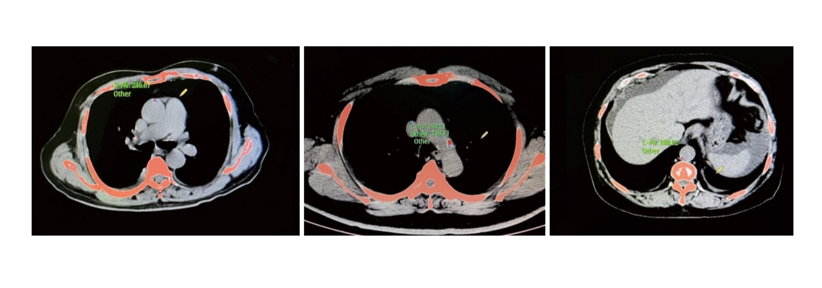 Association between CHA2DS2-VASc score and aortic calcification in patients with atrial fibrillation and its sex-specific predictive value
 Extra-anatomic venous bypass using prosthetic grafts for hemodialysis-related symptomatic central venous stenosis: a single-center retrospective study
 Establishment and preliminary validation of a porcine femoral artery chronic total occlusion-like lesion model induced by combined CaCl2 and FeCl3 treatment
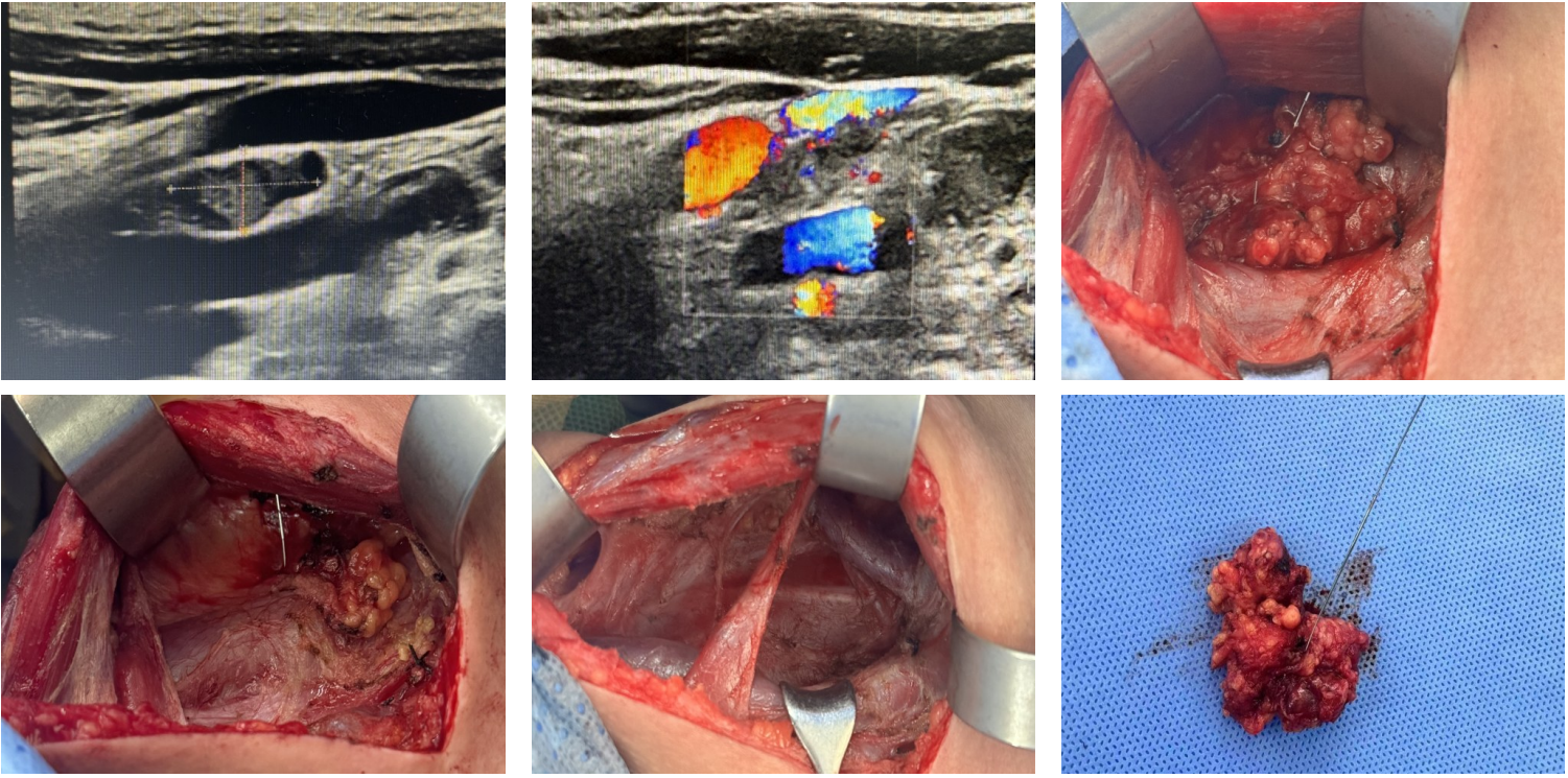 Clinical value of ultrasound-guided guidewire localization in reoperation for recurrent metastatic lymph nodes after thyroid cancer surgery
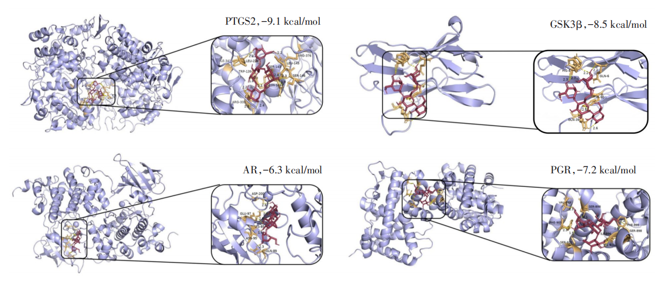 Anti-tumor effects and potential mechanisms of oyster glycogen in papillary thyroid carcinoma: a network pharmacology and in vitro experimental study
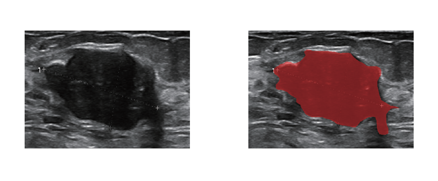 Clinical value of a machine learning model integrating ultrasound radiomics and clinical features for predicting pathological complete response to neoadjuvant therapy in HER2-positive breast cancer
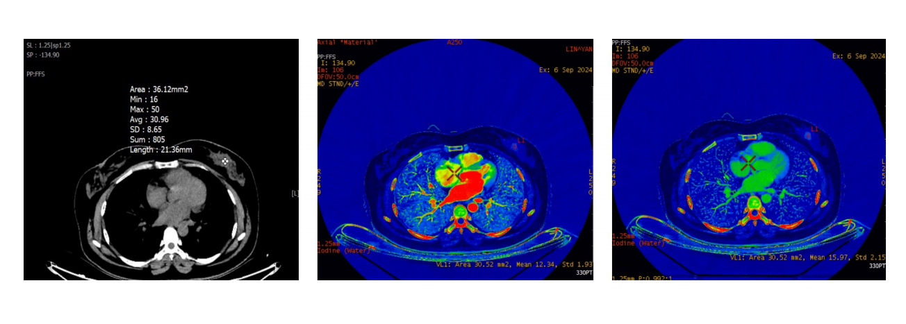 Development of a prediction model for high Ki-67 expression in invasive breast cancer based on dual-energy CT quantitative parameters
 Guidelines for the diagnosis and treatment of primary liver cancer (2026 edition)
- Current Issue
- Published Ahead-of-Print
- Virtual Issues
- Previous Issues
-
Hepatobiliary Surgery Committee of Hunan Medical Association, Hepatobiliary Enhanced Recovery After Surgery Group, Hepatobiliary Surgery Committee of Hunan Medical Association, Enhanced Recovery After Surgery Committee of Hunan Health Management Association, Hepatobiliary Surgery Committee of Hunan Association for International Medical Exchange, Promotion, Hunan Hepatobiliary, Pancreatic Surgery Alliance, Research Center for Hepatobiliary, Pancreatic Diseases, Furong Laboratory
2026,35(8):1481-1507, DOI: 10.7659/j.issn.1005-6947.260452
Abstract:
To further standardize the implementation of enhanced recovery after surgery (ERAS) in hepatobiliary and pancreatic surgery, the 2022 edition of the Hunan expert consensus on the clinical pathway of enhanced recovery after surgery for hepatobiliary and pancreatic diseases was systematically updated based on implementation experience, newly available national and international evidence from 2022 to 2026, multicenter clinical practice data from Hunan Province, and the resources available at healthcare institutions of different levels. The updated consensus comprises 60 recommendations covering three domains: preoperative, intraoperative, postoperative. Major updates and refinements include frailty assessment and intervention, perioperative sleep management, goal-directed blood pressure and fluid management, standardized intraoperative antimicrobial prophylaxis, risk-stratified management of tubes and drains, post-discharge follow-up, and standardized data collection and artificial intelligence-assisted prediction of postoperative complications. This consensus aims to provide healthcare institutions at different levels with a stratified, measurable, and continuously updatable ERAS clinical pathway for hepatobiliary and pancreatic surgery.
-
Chinese Society for Metabolic, Bariatric Surgery, Beijing Metabolic & Bariatric Doctor Association
2026,35(8):1508-1519, DOI: 10.7659/j.issn.1005-6947.260361
Abstract:
Intrathoracic gastric migration (ITGM) is a common complication following metabolic bariatric surgery, occurring after various procedures, with the highest reported incidence after sleeve gastrectomy. The pathogenesis of ITGM is multifactorial, including intraoperative anatomical disruption, postoperative weakening of gastric fixation structures, and thoracoabdominal pressure gradients. Patients may be asymptomatic or present with symptoms related to gastroesophageal reflux disease. Currently, there is no unified diagnostic criteria or standardized management strategy for ITGM. To improve the diagnosis and treatment of ITGM after metabolic bariatric surgery in China, the Chinese Society for Metabolic and Bariatric Surgery and Beijing Metabolic & Bariatric Doctor Association convened a panel of 117 experts nationwide to develop a consensus based on the latest evidence and clinical experience. The consensus addresses 16 key issues, including diagnostic methods, monitoring strategies, indications for surgical intervention, and specific management protocols for ITGM following different metabolic bariatric procedures. This consensus provides evidence-informed recommendations for the diagnosis and management of ITGM after metabolic bariatric surgery in China. Further high-quality prospective studies are needed to strengthen the evidence base and optimize future recommendations.
-
2026,35(8):1520-1528, DOI: 10.7659/j.issn.1005-6947.260449
Abstract:
Biliopancreatic diseases share common embryonic origins, close anatomical relationships, interconnected physiological functions, and similar disease spectra, with substantial similarities in their pathogenesis, clinical manifestations, diagnosis, and treatment. Therefore, an integrated perspective is important for identifying the underlying causes, standardizing clinical management, and optimizing therapeutic strategies for biliopancreatic diseases. However, current clinical practice is still characterized by excessive subspecialization and fragmentation, insufficient holistic thinking, overreliance on imaging findings with inadequate attention to pathological evidence, and insufficient recognition of the function and preservation of the sphincter of Oddi. For challenging conditions, including unexplained biliopancreatic duct dilatation, pancreatic-segment choledochal cysts, distal bile duct strictures, and sphincter of Oddi dysfunction, comprehensive assessment of the biliopancreatic system should be emphasized. Multidisciplinary team management and systematic follow-up should be incorporated into clinical decision-making to reduce missed diagnoses, inappropriate treatment, and overtreatment. Meanwhile, research from an integrated perspective should focus on the patterns of biliopancreatic duct confluence, the pathogenesis and precision management of biliopancreatic malignancies, and the basic and clinical characteristics of sphincter of Oddi function, thereby further improving the scientific, standardized, and effective management of biliopancreatic diseases.
- 1
- 2
-
CHEN Guanyu, WANG Xitao, TAN Jian, LI Yunfeng
2026,35(8):1529-1541, DOI: 10.7659/j.issn.1005-6947.260379
Abstract:
Benign biliary stricture following bilioenteric anastomosis is an important late complication of biliary reconstruction and may result in cholestasis, recurrent cholangitis, intrahepatic bile duct stones, and secondary hepatic dysfunction. For patients with failed or unsuitable endoscopic or percutaneous interventions, particularly those with severe cicatricial strictures, high hilar or multifocal biliary involvement, or complex intrahepatic biliary disease, redo bilioenteric anastomosis remains an important option for restoring durable biliary drainage. With its three-dimensional magnified visualization, wristed instruments, and enhanced suturing capability, robotic surgery provides a potential minimally invasive approach for complex biliary reconstruction. Based on the relevant literature and our institutional experience, we systematically summarize the key aspects of robot-assisted redo bilioenteric anastomosis for benign biliary strictures after bilioenteric anastomosis, including patient selection, preoperative assessment, trocar placement, adhesiolysis, hilar dissection, bile duct incision and ductoplasty, basin-like bilioenteric drainage reconstruction, perioperative management, and postoperative follow-up. Particular attention is given to technical principles for reducing anastomotic tension, preserving bile duct vascularity, creating a wide and effective drainage surface, and achieving precise mucosa-to-mucosa apposition during complex biliary reconstruction. Robot-assisted redo bilioenteric anastomosis may provide a feasible minimally invasive reconstructive option for selected patients with complex benign biliary strictures after bilioenteric anastomosis. Its application should, however, be based on strict patient selection, comprehensive preoperative assessment, and adequate expertise in complex biliary reconstruction, while its long-term efficacy and potential advantages over other surgical approaches require further validation in high-quality clinical studies.
- 1
- 2
- 3
- 4
- 5
-
WANG Jiang, SUN Wensheng, XU Zhuangzhi, YU Xiutao
2026,35(8):1542-1552, DOI: 10.7659/j.issn.1005-6947.250711
Abstract:
Background and Aims Lymph node metastasis (LNM) is a major determinant of prognosis in patients with gallbladder carcinoma (GBC). Accurate preoperative assessment of LNM is essential for individualized treatment planning. This study investigated the predictive value of the inflammatory burden index (IBI) combined with hypoxia-inducible factor 1α (HIF-1α) for LNM and evaluated their interaction in patients with GBC.Methods Clinical data from 106 patients with pathologically confirmed GBC treated at Linyi Central Hospital between January 2020 and March 2025 were retrospectively analyzed. Patients were divided into an LNM group (n=44) and a non-LNM group (n=62) according to postoperative pathological findings. Differences in IBI and HIF-1α levels were compared between groups. Independent risk factors for LNM were identified using multivariate Logistic regression analysis. Receiver operating characteristic curve analysis was used to evaluate predictive performance. Regression discontinuity design (RDD) was applied to determine threshold effects. Additive and multiplicative interaction analyses were performed to assess the synergistic effects of IBI and HIF-1α. Subgroup analyses were conducted according to tumor differentiation and T stage.Results Patients with LNM exhibited significantly higher IBI and HIF-1α levels than those without LNM (both P<0.001). Multivariate Logistic regression analysis identified T stage, IBI, and HIF-1α as independent risk factors for LNM (all P<0.05). The combined model demonstrated good predictive performance with an area under the curve of 0.847 (95% CI=0.782-0.902), outperforming either marker alone. RDD analysis identified significant thresholds at IBI<33.21 and HIF-1α<9.65 pg/mL, below which the risk of LNM decreased significantly. Significant synergistic interactions between IBI and HIF-1α were observed on both additive and multiplicative scales. Subgroup analyses showed that the combined model maintained superior predictive performance across different differentiation grades and T stages, particularly in poorly differentiated tumors and T3-T4 stage disease.Conclusion Preoperative IBI and HIF-1α are independent predictors of LNM in GBC. Their combined assessment significantly improves predictive accuracy and demonstrates synergistic effects, providing a useful tool for preoperative risk stratification and individualized therapeutic decision-making for GBC patients.
- 1
- 2
- 3
- 4
-
JIA Yapeng, WANG Yu, LI Yongkun
2026,35(8):1553-1562, DOI: 10.7659/j.issn.1005-6947.260399
Abstract:
Background and Aims Intrahepatic cholangiocarcinoma (ICC) is a highly aggressive primary liver malignancy, and radical resection remains the main treatment option for achieving long-term survival. Laparoscopic anatomical hepatectomy and regional lymph node dissection require precise identification of hepatic anatomy and accurate surgical manipulation. Three-dimensional visualization technology facilitates individualized preoperative surgical planning, whereas indocyanine green (ICG) fluorescence navigation provides real-time intraoperative imaging. This study investigated the clinical value of combining ICG fluorescence navigation with three-dimensional visualization technology in laparoscopic anatomical hepatectomy and regional lymph node dissection for ICC.Methods This prospective, single-center, open-label, randomized controlled study included 122 patients who underwent laparoscopic radical resection for ICC between April 2023 and April 2025. Patients were randomly assigned at a 1∶1 ratio to a conventional group or a combined group, with 61 patients in each group. Patients in the conventional group underwent laparoscopic radical resection alone, whereas those in the combined group received ICG fluorescence navigation combined with three-dimensional visualization in addition to the conventional procedure. Intraoperative parameters, short-term postoperative recovery, postoperative complications, tumor recurrence, and survival outcomes were compared between the groups. The primary endpoints were evaluated according to the intention-to-treat (ITT) principle, with per-protocol set (PPS) analysis performed as a supportive analysis.Results In the PPS analysis, the combined group had shorter hepatic portal occlusion time, less intraoperative blood loss, and a lower intraoperative transfusion rate than the conventional group (all P<0.05). The rates of compliant anatomical hepatectomy, adequate lymph node dissection, and R0 resection were higher in the combined group (all P<0.05). The combined group also had a greater total number of dissected lymph nodes and positive lymph nodes (both P<0.05), and eight satellite lesions were additionally detected in the combined group compared with none in the conventional group. The drainage tube removal time and hospital stay were shorter, and postoperative day 3 levels of alanine aminotransferase, aspartate aminotransferase, and total bilirubin were lower in the combined group (all P<0.05). The overall incidence of postoperative complications was lower in the combined group than in the conventional group (5.36% vs. 19.64%, P=0.022). The 1-year regional lymph node recurrence rate was also lower in the combined group (3.57% vs. 14.29%, P=0.047), whereas no significant differences were observed in 1-year intrahepatic recurrence or distant metastasis rates (both P>0.05). Survival analysis showed that the combined group had longer progression-free survival than the conventional group (median, 30 months vs. 26 months; P=0.037), while overall survival did not differ significantly between the groups (median, 32 months vs. 27 months; P=0.215). The ITT analysis yielded results generally consistent with those of the PPS analysis.Conclusion The combination of ICG fluorescence navigation and three-dimensional visualization technology in laparoscopic anatomical hepatectomy and regional lymph node dissection for ICC may improve the precision of anatomical localization and lymph node dissection, reduce intraoperative blood loss and hepatic portal occlusion time, and facilitate postoperative recovery. It may also reduce regional lymph node recurrence and prolong progression-free survival; however, its effect on long-term overall survival requires confirmation in larger multicenter studies.
- 1
-
LIANG Yucen, ZHANG Honglong, WANG Guoqiang, QIN Wenke, WANG Hongtao
2026,35(8):1563-1572, DOI: 10.7659/j.issn.1005-6947.260385
Abstract:
Background and Aims Acute obstructive suppurative cholangitis (AOSC) is a severe biliary tract infection characterized by rapid onset and progression, for which timely relief of biliary obstruction is essential. With the development of laparoscopic techniques, laparoscopic common bile duct exploration (LCBDE) has become an important surgical approach for common bile duct stones. After stone extraction, primary suture (PS) or T-tube drainage (TD) can be used to manage the common bile duct incision; however, the clinical efficacy and safety of these two approaches in patients with AOSC remain incompletely defined. In particular, postoperative bile leakage is an important complication of LCBDE-PS that may adversely affect postoperative recovery. Therefore, identification of its risk factors and risk stratification are of clinical importance. This study aimed to compare the clinical efficacy of LCBDE plus PS and LCBDE plus TD in patients with AOSC, identify factors associated with postoperative bile leakage after LCBDE plus PS, and develop a prediction model for postoperative bile leakage to provide a reference for surgical decision-making and postoperative risk assessment.Methods The clinical data of 186 patients with AOSC admitted to Wuwei People's Hospital from January 2023 to December 2025 were retrospectively analyzed. Among them, 96 patients underwent LCBDE with primary suture (LCBDE-PS group), while 90 underwent LCBDE with T-tube drainage (LCBDE-TD group). Perioperative outcomes, postoperative complications, and stone recurrence at 5 months after surgery were compared between the two groups. Patients in the LCBDE-PS group were further divided into a bile leakage group (n=11) and a non-bile leakage group (n=85) according to the occurrence of postoperative bile leakage. Clinical and laboratory parameters were compared between the two groups. Multivariate Logistic regression analysis was performed to identify independent factors associated with postoperative bile leakage after LCBDE-PS, and a nomogram prediction model was subsequently constructed. The Hosmer-Lemeshow goodness-of-fit test, receiver operating characteristic (ROC) curve, and decision curve analysis (DCA) were used to evaluate the model's goodness of fit, discriminative ability, and clinical net benefit, respectively.Results Compared with the LCBDE-TD group, the LCBDE-PS group had shorter operative time, less intraoperative blood loss, shorter drainage-tube indwelling time, lower total abdominal drainage volume, and shorter postoperative hospital stay (all P<0.05). No significant differences were observed between the two groups in the incidence of postoperative complications or stone recurrence during follow-up (all P>0.05). Within the LCBDE-PS group, patients with bile leakage had higher preoperative direct bilirubin levels and longer operative times, but lower preoperative albumin levels and smaller common bile duct diameters than those without bile leakage (all P<0.05). Multivariate Logistic regression analysis showed that a larger common bile duct diameter (OR=0.658, 95% CI=0.505-0.857, P=0.002) and higher preoperative albumin level (OR=0.719, 95% CI=0.550-0.940, P=0.016) were associated with a lower risk of postoperative bile leakage, whereas higher preoperative direct bilirubin level (OR=1.435, 95% CI=1.161-1.773, P=0.001) and longer operative time (OR=2.037, 95% CI=1.092-3.799, P=0.025) were associated with an increased risk. The nomogram incorporating these four factors showed good model fit. The area under the ROC curve was 0.949 (95% CI=0.901-0.997), with a sensitivity of 90.91% and specificity of 89.41%. DCA indicated a positive net clinical benefit of the model across a threshold probability range of 0.03-0.96.Conclusion Compared with LCBDE plus TD, LCBDE plus PS may provide favorable perioperative outcomes for patients with AOSC without increasing postoperative complications or short-term stone recurrence. Common bile duct diameter, preoperative albumin level, preoperative direct bilirubin level, and operative time were associated with postoperative bile leakage after LCBDE plus PS. The nomogram based on these factors showed good discriminative performance and may assist in postoperative risk stratification for bile leakage.
- 1
- 2
-
QIU Riwei, WEI Jiang, XIAO Weidong
2026,35(8):1573-1581, DOI: 10.7659/j.issn.1005-6947.260139
Abstract:
Background and Aims Patients with a history of upper abdominal surgery often present with increased difficulty and surgical risk during laparoscopic common bile duct exploration (LCBDE) because of intra-abdominal adhesions, altered anatomical structures, and difficulties in exposing the biliary tract. Indocyanine green (ICG) fluorescence imaging may facilitate intraoperative bile duct localization; however, its clinical utility in these patients remains unclear. This study aimed to evaluate the clinical utility of ICG fluorescence imaging-assisted laparoscopic common bile duct exploration in patients with common bile duct stones and a history of upper abdominal surgery.Methods The clinical data of patients with common bile duct stones and a history of upper abdominal surgery who underwent LCBDE at the First Affiliated Hospital of Nanchang University between May 2019 and June 2024 were retrospectively analyzed. Patients were divided into an ICG group and a non-ICG group according to whether ICG fluorescence imaging was used during operation. The primary outcomes assessed included bile duct identification time, operative time, intraoperative blood loss, intraoperative complications, conversion to open surgery, bile duct management method, postoperative length of stay, postoperative complications, residual stones, and stone recurrence.Results A total of 178 patients were included, comprising 64 in the ICG group and 114 in the non-ICG group. No statistically significant differences in baseline characteristics were observed between the two groups. Successful bile duct fluorescence visualization was achieved in 58 of 64 patients in the ICG group (90.6%). Compared with the non-ICG group, the ICG group had shorter median bile duct identification time, shorter operative time, and less intraoperative blood loss [20.00 (15.00-25.00) min vs. 50.00 (31.25-60.00) min, P<0.001; 157.50 (130.00-210.00) min vs. 190.00 (146.25-260.00) min, P=0.005; and 50.00 (50.00-100.00) mL vs. 100.00 (50.00-200.00) mL, P=0.002, respectively]. No statistically significant differences were observed in intraoperative complications, conversion to open surgery, bile duct management method, postoperative length of stay, postoperative complications, residual stone rate, or stone recurrence rate between the two groups (all P>0.05).Conclusion In patients with common bile duct stones and a history of upper abdominal surgery, ICG fluorescence imaging-assisted LCBDE was associated with shorter bile duct identification and operative times and less intraoperative blood loss. ICG fluorescence imaging may facilitate bile duct localization and intraoperative manipulation in patients with complex abdominal anatomy. Its effects on postoperative and long-term outcomes warrant further investigation.
-
DU Guohui, MENG Lingzhan, LI Hu, LIU Jia, WANG Xuexin, ZHAI Yongxiang, DAN Hanlei, YAN Jin, SHAO Yanling, ZHU Zhenyu
2026,35(8):1582-1582, DOI: 10.7659/j.issn.1005-6947.260116
Abstract:
Background and Aims Long-standing hepatolithiasis with recurrent biliary inflammation may lead to liver lobe atrophy-hypertrophy syndrome. In some patients, marked atrophy of both hepatic lobes is accompanied by compensatory enlargement of the caudate lobe, making the extent of hepatectomy and accurate assessment of the future liver remnant challenging. We report a case of hepatolithiasis complicated by liver lobe atrophy-hypertrophy syndrome treated with caudate lobe-sparing subtotal hepatectomy and review the relevant literature to evaluate the indications, key technical considerations, and clinical applicability of this procedure.Methods The clinical data of a 66-year-old patient with hepatolithiasis complicated by liver lobe atrophy-hypertrophy syndrome who was admitted to the Fifth Medical Center of Chinese PLA General Hospital in March 2025 were retrospectively analyzed. Three-dimensional visualization was used preoperatively to assess hepatic morphology, caudate lobe volume, and the anatomical relationships of its vascular and biliary structures. Surgical planning was performed based on these findings and hepatic functional reserve. Caudate lobe-sparing subtotal hepatectomy combined with common bile duct exploration, stone extraction, and T-tube drainage was subsequently performed with the assistance of choledochoscopy and intraoperative ultrasonography. Relevant cases reported in the literature were also reviewed.Results Imaging revealed marked atrophy of the left and right hepatic lobes, compensatory enlargement of the caudate lobe, and multiple intrahepatic biliary stones with intrahepatic bile duct dilatation. The caudate lobe had preserved blood supply, venous drainage, and biliary drainage. Three-dimensional reconstruction showed a caudate lobe volume of 948 mL, corresponding to approximately 84% of the standard liver volume after resection. Caudate lobe-sparing subtotal hepatectomy combined with common bile duct exploration, stone extraction, and T-tube drainage was successfully performed. The operative time was 420 min, with estimated blood loss of approximately 800 mL and no blood transfusion. Liver function recovered well postoperatively, with no severe complications, and the patient was discharged on postoperative day 12. Histopathological examination unexpectedly revealed intrahepatic cholangiocarcinoma, large-duct type, with negative resection margins. At 2 months postoperatively, no definite residual stones or tumor recurrence was detected. Choledochoscopy at 4 months showed no residual biliary stones, and the T-tube was removed. During the 8-month follow-up, the patient remained in good general condition, with no definite evidence of recurrent stones or tumor on imaging.Conclusion In carefully selected patients with hepatolithiasis complicated by liver lobe atrophy-hypertrophy syndrome, caudate lobe-sparing subtotal hepatectomy is feasible after comprehensive assessment of hepatic functional reserve, future liver remnant volume, and the vascular, venous, and biliary drainage of the caudate lobe. Three-dimensional visualization, choledochoscopy, and intraoperative ultrasonography may facilitate precise preoperative planning and intraoperative identification and preservation of critical structures. However, this technically demanding procedure requires strict patient selection, and long-term complications such as intrahepatic cholangiocarcinoma should be carefully considered.
- 1
- 2
- 3
- 4
- 5
- 6
-
PENG Baiming, ZHANG Xiaoyu, FENG Longbao, GAO Yuanhui, ZHANG Jianquan, XIANG Yang
2026,35(8):1591-1606, DOI: 10.7659/j.issn.1005-6947.250627
Abstract:
Background and Aims Biliary stricture remains challenging to treat because of limitations related to stent mechanical performance, restenosis, and postoperative monitoring. This study aimed to develop a 3D-printed ultrasmall superparamagnetic iron oxide (USPIO)/epigallocatechin gallate (EGCG) functionalized bilayer hydrogel biliary stent integrating mechanical support, local drug delivery, and MRI visibility, and to evaluate its physicochemical and in vitro biological properties.Methods Polycaprolactone methacrylate (PCLMA) was used to construct the mechanically supportive inner layer, while gelatin methacryloyl (GelMA) was used as the bioactive outer layer. Mesoporous silica nanoparticles (MSN) loaded with EGCG and USPIO were incorporated into the hydrogel system. A PCLMA/GelMA/MSN@USPIO@EGCG bilayer hydrogel biliary stent was fabricated by 3D printing and photocrosslinking. The structural and physicochemical properties of the materials were characterized using transmission electron microscopy, scanning electron microscopy, Fourier-transform infrared spectroscopy, proton nuclear magnetic resonance spectroscopy, dynamic light scattering, and rheological analysis. In vitro degradation and EGCG release were evaluated, and MRI was used to assess the imaging capability conferred by USPIO. The cytocompatibility, cell proliferation, reactive oxygen species (ROS), mitochondrial membrane potential, and cellular senescence of human adipose-derived mesenchymal stem cells (hADSC) were evaluated. Hemolysis and coagulation assays were performed to assess hemocompatibility.Results The prepared MSN and composite nanoparticles exhibited relatively uniform spherical morphology and good dispersion, while the PCLMA/GelMA/MSN@USPIO@EGCG stent showed a regular porous grid-like structure. The PCLMA inner layer exhibited high compressive strength, whereas the GelMA-based composite hydrogel showed favorable viscoelastic properties. The composite stent exhibited controllable degradation and sustained EGCG release in vitro. USPIO incorporation conferred a distinct T2-weighted negative MRI contrast effect, with a strong linear relationship between the relaxation rate and iron concentration. All stent groups showed good cytocompatibility and supported hADSCs adhesion and spreading. EGCG incorporation further enhanced cell proliferation, reduced oxidative stress, and helped maintain mitochondrial membrane potential. The composite stent caused no obvious hemolytic or coagulation abnormalities over the tested concentration and incubation ranges.Conclusion A 3D-printed bilayer hydrogel biliary stent with mechanical support, sustained EGCG release, and MRI visibility was successfully developed. The stent exhibited favorable biodegradability, cytocompatibility, and cytoprotective properties in vitro, providing a potential biomaterial strategy for local microenvironment modulation and imaging-assisted monitoring of biliary strictures. Its in vivo safety and anti-restenotic efficacy require further investigation.
- 1
- 2
- 3
- 4
- 5
- 6
- 7
- 8
- 9
- 10
- 11
- 12
- 13
- 14
-
LONG Fei, LIANG Ni, ZHENG Xunri, YE Yongxin, CHEN Miao
2026,35(8):1607-1619, DOI: 10.7659/j.issn.1005-6947.260287
Abstract:
Background and Aims Ferroptosis is an iron-dependent form of regulated cell death characterized by lipid peroxidation and has potential applications in colorectal cancer (CRC) therapy. Circular RNAs (circRNAs) can regulate tumor progression through competing endogenous RNA (ceRNA) mechanisms; however, their roles and regulatory mechanisms in CRC ferroptosis remain incompletely understood. This study aimed to identify dysregulated circRNAs in CRC, investigate the effects of hsa_circ_0001819 (circ1819) on ferroptosis and malignant phenotypes of CRC cells, and preliminarily explore its potential molecular mechanism.Methods CircRNA microarray datasets GSE223001 and GSE205643 were integrated to identify differentially expressed circRNAs, with GSE126094 used for validation. The expression of circ1819 in CRC tissues and cell lines was examined by qRT-PCR. RNase R digestion, actinomycin D treatment, and nuclear-cytoplasmic fractionation assays were performed to characterize its circular structure, stability, and subcellular localization. Following siRNA-mediated circ1819 knockdown, cell viability, lipid reactive oxygen species (ROS), malondialdehyde (MDA), and ferroptosis-related proteins were assessed in the presence of the ferroptosis inducer RSL3 and inhibitor Liproxstatin-1 (Lip-1). CCK-8, colony formation, and Transwell assays were used to evaluate cell proliferation and migration. AGO2 RNA immunoprecipitation (AGO2-RIP), RNA pulldown, and bioinformatics analyses were performed to investigate the potential ceRNA regulatory mechanism of circ1819.Results Circ1819 was significantly upregulated in CRC tissues and cell lines, and its expression was associated with tumor diameter and T stage. Circ1819 exhibited typical characteristics of a circular RNA and was predominantly localized in the cytoplasm. Circ1819 knockdown significantly increased the sensitivity of CRC cells to RSL3-induced ferroptosis, as evidenced by reduced cell viability and increased lipid ROS and MDA levels; these effects were partially reversed by Lip-1. Circ1819 knockdown significantly reduced GPX4 mRNA and protein expression, whereas SLC7A11 and ACSL4 expression remained unchanged. Moreover, circ1819 knockdown significantly inhibited CRC cell proliferation, colony formation, and migration. AGO2-RIP and RNA pulldown assays suggested interactions between circ1819 and miR-618 or miR-1231. Combined with target prediction and enrichment analyses, a potential circ1819-centered circRNA-miRNA-mRNA regulatory network was constructed.Conclusion Circ1819 is aberrantly upregulated in CRC, and its knockdown enhances ferroptosis sensitivity while suppressing proliferation and migration of CRC cells. These effects may be associated with ceRNA regulation involving miR-618 and miR-1231 and alterations in GPX4 expression. Circ1819 may represent a potential regulatory link between malignant progression and ferroptosis resistance in CRC, although its downstream targets and causal regulatory relationships require further validation.
- 1
- 2
- 3
- 4
- 5
-
2026,35(8):1620-1631, DOI: 10.7659/j.issn.1005-6947.250327
Abstract:
Background and Aims Hypoxia is a key feature of the tumor microenvironment of hepatocellular carcinoma (HCC) and contributes to tumor progression through hypoxia-inducible factor (HIF)-mediated regulation of multiple tumor-promoting genes. This study aimed to investigate the relationship between ring finger protein 24 (RNF24) and hypoxia/HIF-1α, evaluate RNF24 expression and its prognostic significance in HCC, and explore its effects on the malignant biological behaviors of HCC cells under hypoxic conditions and the underlying mechanisms.Methods The correlation between RNF24 and HIF-1α expression and the clinicopathological significance of RNF24 were analyzed using transcriptomic data from the TCGA-HCC cohort. RNF24 protein expression was examined by immunohistochemistry in tumor and paired adjacent tissues from 35 patients with HCC. Kaplan-Meier analysis and univariate and multivariate Cox regression analyses were performed to evaluate the prognostic significance of RNF24. Hypoxia was simulated by culturing cells under 1% O2, HIF-1α knockdown and DMOG treatment were used to investigate the regulation of RNF24 by HIF-1α. RNF24 knockdown, CCK-8, and Transwell assays were performed to assess the effects of RNF24 on HCC cell proliferation, migration, and invasion under hypoxic conditions. Gene set enrichment analysis (GSEA), correlation analysis, qRT-PCR, and Western blot were further performed to investigate the potential molecular mechanisms underlying the biological effects of RNF24.Results TCGA analysis showed that RNF24 expression was positively correlated with HIF-1α expression. Hypoxia and DMOG treatment significantly increased RNF24 expression in HCC cells, whereas HIF-1α knockdown markedly attenuated hypoxia-induced RNF24 upregulation, suggesting that RNF24 is regulated by HIF-1α. RNF24 expression was significantly higher in HCC tissues than in corresponding adjacent tissues in both the TCGA cohort and the clinical cohort. High RNF24 expression was associated with aggressive clinicopathological features, including advanced TNM stage, higher T stage, and vascular invasion. Kaplan-Meier analysis showed that patients with high RNF24 expression had poorer overall survival rate and disease-free survival rate. Multivariate Cox regression analysis identified high RNF24 expression as an independent risk factor for poor prognosis in HCC (HR=1.407, 95% CI=1.058-1.872, P=0.019). Functional assays demonstrated that hypoxia promoted HCC cell proliferation, migration, and invasion, whereas RNF24 knockdown markedly attenuated these effects. GSEA showed that high RNF24 expression was associated with enrichment of the mTOR signaling pathway. Moreover, RNF24 knockdown reduced the expression of HIF-1α and its target genes, including VEGFA, TWIST, MMP2, and GLUT1.Conclusion RNF24 is a hypoxia-responsive gene regulated by HIF-1α and is highly expressed in HCC tissues. High RNF24 expression is associated with aggressive clinicopathological characteristics and poor prognosis. RNF24 contributes to hypoxia-induced HCC cell proliferation, migration, and invasion and may exert its tumor-promoting effects by enhancing HIF-1α and its downstream target gene expression. RNF24 may serve as a potential molecular biomarker and therapeutic target for HCC progression.
- 1
- 2
- 3
- 4
- 5
-
WANG Kangtao, ALI Muhammad Kashif, PAN Baihong, OUYANG Yang
2026,35(8):1632-1642, DOI: 10.7659/j.issn.1005-6947.260360
Abstract:
Background and Aims Venous thromboembolism (VTE) is an important complication after spine surgery. Although the Caprini score is widely used for perioperative VTE risk stratification, it has limited ability to capture early postoperative information related to coagulation and fibrinolysis, nutritional status, and surgical burden. This study aimed to evaluate the incremental predictive value of early postoperative clinical information beyond the postoperative Caprini score and to develop and internally validate models for predicting in-hospital VTE after spine surgery.Methods This single-center retrospective cohort study included 1 508 adult patients who underwent spine surgery at Xiangya Hospital, Central South University, in 2024. The primary outcome was overall VTE occurring during postoperative hospitalization. Postoperative laboratory variables were defined as the first measurements obtained within 24-48 h after surgery; for patients who developed VTE, only measurements obtained before VTE diagnosis were used. Three Logistic regression models were developed. Model A included the postoperative Caprini score alone. Model B additionally included age, albumin, fibrin degradation products (FDP), operation duration, intraoperative blood loss, and transfused red blood cell units. Model C included the postoperative Caprini score, age, albumin, and FDP. Internal validation was performed using 1 000 bootstrap resamples and stratified 10-fold cross-validation. Model discrimination, calibration, and clinical utility were assessed using the area under the receiver operating characteristic curve (AUC), calibration analysis, and decision curve analysis. A preoperative Caprini score model, a sensitivity analysis excluding FDP, and an auxiliary LASSO analysis were also performed. Based on the validated models B and C, online web-based risk assessment tools were developed to enable individualized VTE risk calculation and visual assessment.Results Among the 1 508 patients, 156 developed VTE, corresponding to an incidence of 10.34%. After bootstrap optimism correction, the AUC of models A, B, and C were 0.706, 0.824, and 0.821, respectively. The corresponding out-of-fold AUC from 10-fold cross-validation were 0.699 (95% CI=0.656-0.742), 0.824 (95% CI=0.792-0.855), and 0.820 (95% CI=0.790-0.851). Models B and C demonstrated significantly better discrimination than Model A (both P<0.001), whereas no significant difference was observed between models B and C (P=0.494). The calibration slopes of models B and C were 0.954 and 0.975, respectively. Decision curve analysis showed that models B and C generally provided greater net benefit than model A across threshold probabilities of approximately 0.02-0.30. After excluding FDP, the out-of-fold AUC of the enhanced model was 0.795; paired DeLong comparison with model B yielded an AUC difference of 0.028 (P=0.006), indicating an incremental predictive contribution of FDP. Online web-based risk assessment tools were developed based on the validated models B and C, enabling individualized VTE risk calculation and visual assessment.Conclusion The postoperative Caprini score provides baseline risk information for in-hospital VTE after spine surgery but has limited discrimination when used alone. Incorporating age, albumin, FDP, and surgery-related information further improves risk discrimination. The simplified model maintained predictive performance comparable to that of the full enhanced model while using fewer predictors, supporting its potential use for postoperative VTE risk stratification and online assessment. External validation in independent multicenter cohorts is required before broader clinical application.
- 1
- 2
-
YUAN Ting, YAN An, DENG Xiaoxian, YANG Lihong
2026,35(8):1643-1653, DOI: 10.7659/j.issn.1005-6947.260295
Abstract:
Background and Aims Pancreatic cancer (PC) has a poor prognosis, and etiological evidence regarding potentially modifiable risk factors remains limited. Blood metals are involved in oxidative stress, metabolic regulation, and immune responses, but their causal relationships with PC risk remain unclear. This study aimed to systematically evaluate the potential causal associations between 42 blood metal traits and PC risk using a multi-ancestry Mendelian randomization (MR) framework.Methods Genome-wide association study (GWAS) summary statistics for 42 blood metal traits in East Asian populations were used as exposure data. Two-sample MR analyses were conducted using three PC GWAS datasets, including two East Asian datasets and one European dataset. The inverse-variance weighted (IVW) method was used as the primary analysis, with MR-Egger, weighted median, and weighted mode as complementary approaches. IVW estimates for all 42 metals across the three PC GWAS datasets were subsequently combined using random-effects meta-analysis. Multiple testing was addressed using Bonferroni and Benjamini-Hochberg false discovery rate (FDR) correction. Heterogeneity, horizontal pleiotropy, outliers, leave-one-out analyses, and Steiger directionality testing were performed to assess robustness. MRlap was further used to evaluate the potential impact of sample overlap between the East Asian exposure and outcome GWAS datasets.Results Plasma iron showed directionally consistent positive associations with PC risk across all three PC GWAS datasets and reached nominal statistical significance in each dataset. Random-effects meta-analysis demonstrated that higher genetically predicted plasma iron levels were significantly associated with increased PC risk (pooled OR=1.195, 95% CI=1.089-1.311, P=1.73×10⁻⁴). This association remained significant after Bonferroni correction for 42 comparisons (adjusted P=0.007; FDR-adjusted P=0.007), with no significant between-dataset heterogeneity (I2=0%). Plasma copper showed a nominally significant meta-analytic association and remained significant after FDR correction (FDR-adjusted P=0.042), but not after Bonferroni correction (Bonferroni-adjusted P=0.085). Sensitivity analyses revealed no substantial evidence of directional horizontal pleiotropy, instrumental heterogeneity, outlier-driven effects, or single-variant-driven associations for plasma iron, and Steiger testing supported the direction from plasma iron to PC risk.Conclusion This multi-ancestry, multi-metal MR study provides genetic evidence supporting a potential causal association between higher genetically predicted plasma iron levels and increased PC risk. The association remained robust across outcome datasets and after stringent multiple-testing correction, suggesting that iron homeostasis may contribute to the etiology of PC and warranting further mechanistic and risk-intervention studies.
- 1
- 2
-
XIA Weizhi, LI Zhi, LUO Shayang, LI Liya, MA Yixuan, WANG Shouman, LIU Ruijie, WANG Kuansong, LI Ge, TANG Haiyun, ZHOU Weibing, HUANG Juan
2026,35(8):1654-1665, DOI: 10.7659/j.issn.1005-6947.260185
Abstract:
Breast cancer may undergo changes in receptor status and histological characteristics during multiple lines of treatment. In rare cases, metaplastic carcinoma may develop, posing challenges for subsequent management. Herein, we report a case of HR-positive/HER2-negative breast cancer that underwent histological transformation to metaplastic squamous cell carcinoma after multiple lines of therapy. A 50-year-old woman was initially diagnosed with grade Ⅱ invasive carcinoma of the right breast, classified as Luminal A and HER2-negative. She underwent surgery, chemotherapy, radiotherapy, and endocrine therapy combined with abemaciclib. Approximately 28 months after surgery, bone metastasis developed. Pathological examination of the sacral metastatic lesion revealed poorly differentiated adenocarcinoma, with 1% ER positivity, PR negativity, HER2(1+), and a programmed death ligand 1 (PD-L1) combined positive score (CPS) of <1. During subsequent treatment, thoracic vertebral metastasis and spinal cord compression developed. Tumor resection and spinal canal decompression were performed, and postoperative pathology indicated metaplastic carcinoma, specifically poorly differentiated squamous cell carcinoma. The tumor was negative for ER, PR, androgen receptor, and HER2, with a Ki-67 positivity rate of approximately 90%; P40, P63, and CK5/6 were positive, and the PD-L1 CPS was approximately 20. The patient subsequently received capecitabine, trastuzumab deruxtecan, toripalimab combined with bevacizumab, and capivasertib combined with vinorelbine. Four multidisciplinary team (MDT) consultations were conducted based on disease progression, pathological findings, molecular testing, and treatment tolerance. Serial pathological assessments demonstrated a gradual change from invasive carcinoma in the primary tumor to poorly differentiated adenocarcinoma in the sacral metastatic lesion and finally to metaplastic carcinoma with squamous differentiation, accompanied by decreased ER and HER2 expression and increased PD-L1 expression. PIK3CA- and PTEN-related alterations were detected in both the primary tumor and the sacral metastatic lesion. The MDT integrated pathological, radiological, molecular, and clinical information to dynamically adjust the treatment strategy. During treatment with toripalimab combined with bevacizumab, imaging evaluation indicated stable disease; treatment was subsequently modified because of newly developed hepatic and splenic lesions. Advanced breast cancer may undergo dynamic changes in receptor status and histological characteristics during multiple lines of therapy. Timely re-biopsy of metastatic lesions and reassessment of pathological and molecular features may help identify tumor biological evolution and inform subsequent treatment decisions. Sequential MDT consultations may integrate multidisciplinary information and facilitate individualized management of complex advanced breast cancer.
- 1
- 2
- 3
- 4
- 5
- 6
- 7
-
LIN Kangqiang, JIANG Zhao, YE Lin, WENG Jun, YU Yaqun
2026,35(8):1666-1676, DOI: 10.7659/j.issn.1005-6947.250653
Abstract:
Cholecystectomy is a widely used surgical treatment for benign gallbladder diseases, and its potential long-term health consequences have attracted increasing attention. Epidemiological studies have suggested that cholecystectomy may be associated with increased risks of colorectal cancer, cardiovascular disease, metabolic dysfunction-associated steatotic liver disease, cirrhosis, and hepatocellular carcinoma; however, a causal relationship between cholecystectomy and these long-term outcomes has not been established. In this review, we use the bile acid-gut microbiota-inflammation axis as a framework to summarize the interplay among altered bile acid metabolism, gut microbiota dysbiosis, and chronic low-grade inflammation after cholecystectomy, and further discuss their potential roles in the development of colorectal cancer, atherosclerosis, metabolic dysfunction-associated steatotic liver disease, cirrhosis, and hepatocellular carcinoma. Large population-based cohort studies and meta-analyses are integrated with mechanistic evidence to evaluate the consistency and limitations of current findings, with particular emphasis on reverse causation, residual confounding, and indication bias that may complicate causal inference. Potential interventions targeting bile acid signaling, gut microbiota, and metabolic abnormalities are also summarized, and future research priorities are proposed, including identification of high-risk populations, personalized microbiota-based interventions, and multi-omics validation of underlying mechanisms. Current evidence suggests that cholecystectomy may contribute to the development of multiple long-term diseases through disruption of the bile acid-gut microbiota-inflammation axis, but its independent effects and underlying mechanisms remain to be clarified. In clinical practice, the potential long-term metabolic and microbiota-related consequences should be considered alongside the established benefits of cholecystectomy, and appropriate long-term follow-up should be encouraged.
-
2026,35(8):1677-1681, DOI: 10.7659/j.issn.1005-6947.260080
Abstract:
背景与目的 腹腔镜胆囊切除术(LC)后穿刺孔肿瘤种植较为少见,既往胆囊病理提示轻度非典型增生者术后发生腹壁穿刺孔及腹腔多发腺癌更为罕见。本文报告1例LC术后2年出现腹壁穿刺孔肿块并伴梗阻性黄疸的病例,结合文献探讨其可能发生机制及临床管理要点,为胆囊非典型增生患者的术后随访及LC术中无瘤操作提供参考。方法 回顾性分析1例LC术后2年出现腹壁穿刺孔肿块及梗阻性黄疸患者的临床资料,并结合相关文献进行复习。结果 患者为53岁女性,2年前因结石性胆囊炎于外院行LC,术后病理提示慢性胆囊炎伴部分上皮轻度非典型增生,取出胆囊标本时未使用标本袋。术后2年因反复上腹痛、进行性增大的腹壁包块及黄疸入院。增强CT及MRI提示腹壁肿块、肝内外胆管扩张及胰头旁/肝门部异常淋巴结。ERCP提示胆总管中段受压性梗阻,经鼻胆管引流后,黄疸及肝功能明显改善。腹腔镜探查见原剑突下穿刺孔处腹壁与镰状韧带及肝左外叶致密粘连并呈肿瘤浸润,胆囊床、肝门部及周围组织亦呈肿瘤浸润。术中多部位快速冷冻病理均提示高分化腺癌浸润,最终诊断为腹壁穿刺孔及腹腔多发高分化腺癌,因病变广泛浸润无法行根治性切除。术后予胆道支架置入及对症支持治疗,患者未接受进一步抗肿瘤治疗,电话随访6个月时因肿瘤进展死亡。结论 LC术后患者即使既往胆囊病理仅提示轻度非典型增生,仍应重视后续出现恶性病变的可能。对于术中存在胆囊破损、标本未置入标本袋等潜在肿瘤播散风险者,应严格遵循无瘤操作原则;对于病理提示非典型增生的患者,应结合病理分级及手术情况制定个体化随访策略,以便及时发现异常病变。
- 1
- 2
-
2026,35(8):1682-1688, DOI: 10.7659/j.issn.1005-6947.250716
Abstract:
背景与目的 肌间静脉血栓(IMVT)是下肢深静脉血栓(DVT)的常见类型。关于真实抗凝治疗背景下IMVT患者早期下床活动与血栓进展及短期转归之间的关系,现有临床证据仍相对有限。本研究旨在探讨早期下床活动与IMVT患者血栓进展、血栓消失及出血事件之间的相关性。方法 回顾性分析2024年11月—2025年8月中南大学湘雅医院收治的202例IMVT患者的临床资料。根据IMVT确诊后病历记录中的活动情况,将患者分为卧床组(91例)和活动组(111例)。比较两组患者确诊后15 d内血栓进展情况,并随访3个月,观察血栓消失率及出血事件。采用二元Logistic回归和Cox比例风险回归分析血栓进展的相关因素。结果 卧床组15 d内总体血栓事件发生率高于活动组[27.5%(25/91)vs. 6.3%(7/111),P<0.001];其中,近端DVT发生率分别为7.7%(7/91)和0.9%(1/111)(P=0.028),肺栓塞发生率分别为19.8%(18/91)和5.4%(6/111)(P=0.002)。多因素Logistic回归分析显示,卧床与血栓进展相关(OR=4.39,95% CI=1.681~11.364,P=0.002);Cox比例风险回归分析亦显示,卧床与血栓进展风险升高相关(HR=5.015,95% CI=2.161~11.634,P<0.001)。3个月随访时,活动组血栓消失率高于卧床组[95.5%(106/111)vs. 82.4%(75/91),P=0.002]。两组大出血及轻微出血发生率差异均无统计学意义(均P>0.05)。结论 在本研究接受抗凝治疗的IMVT患者中,卧床与15 d内血栓进展及较低的3个月血栓消失率相关。早期下床活动可能与较好的短期临床转归相关,且未观察到出血事件增加。鉴于本研究为单中心回顾性队列研究,相关结论仍需多中心前瞻性研究进一步验证。
- 1
- 2
- 3
Volume 35,2026 Number 8
GUIDELINE AND CONSENSUS
COMMENTARY


SPECIALIST FORUM


MONOGRAPHIC STUDY


BASIC RESEARCH


CLINICAL RESEARCH


MULTIDISCIPLINARY TREATMENT RESEARCH


REVIEW
BRIEF ARTICLES


-
Abstract:
Background and Aims: Inflammation may contribute to the development and progression of pancreatic cancer; however, conventional observational studies have difficulty distinguishing the potential causal relationships between circulating inflammation-related proteins and pancreatic cancer risk from disease-related secondary changes. This study aimed to systematically investigate the potential causal associations between circulating inflammation-related proteins and pancreatic cancer risk using genetic epidemiological approaches and to prioritize candidate proteins with relatively consistent genetic evidence. Methods: Publicly available genome-wide association study (GWAS) summary statistics were used to perform a two-sample Mendelian randomization (MR) analysis of 91 circulating inflammation-related proteins and pancreatic cancer risk. The inverse-variance weighted (IVW) method was used as the primary analytical approach, complemented by weighted median, MR-Egger regression, and weighted mode analyses for robustness assessment. Candidate proteins meeting the prespecified screening criteria were further evaluated using multivariable Mendelian randomization (MVMR) and Mendelian randomization Bayesian model averaging (MR-BMA) to assess their conditional effects and relative model support. Heterogeneity, horizontal pleiotropy, instrument strength, and statistical power were also evaluated. Results: Univariable MR analysis identified five candidate proteins showing nominal associations with pancreatic cancer risk and consistent effect directions across MR methods. Genetically predicted higher levels of cystatin D, CXCL6, and LIFR were associated with increased pancreatic cancer risk, whereas higher levels of IL-2 and IL-24 were associated with lower risk. In subsequent MVMR analyses, CXCL6 was the only protein that consistently retained a positive association across MVMR-IVW, MVMR-Egger, and MVMR-LASSO models. MR-BMA further showed that CXCL6 had the highest marginal inclusion probability (MIP=0.569), with the empirical P value remaining significant after Benjamini-Hochberg correction (P=0.005). Sensitivity analyses revealed no substantial evidence of heterogeneity or directional horizontal pleiotropy. The minimum single-SNP F statistic and conditional F statistic for CXCL6 were 22.67 and 17.49, respectively, with an estimated statistical power of 70.7%, indicating adequate but limited power to detect modest effects. Conclusion: This study identified five circulating inflammation-related proteins potentially associated with pancreatic cancer risk among 91 candidates. CXCL6 showed relatively consistent genetic evidence across MVMR and MR-BMA analyses and may represent a priority candidate for further investigation of inflammation-related pathways in pancreatic cancer susceptibility. However, the current evidence is insufficient to establish a definitive causal relationship between CXCL6 and pancreatic cancer, and further validation in independent populations and tissue-based and functional studies is warranted.
-
Yuanyuan Wu, Huini Liu, An Yan, Hongwei Zhu, XU Long
Abstract:
Background and Aims: Dietary factors may contribute to the development of pancreatic cancer, but conventional observational studies are susceptible to confounding, measurement error, and reverse causation. Food liking is an upstream behavioral phenotype that influences long-term dietary behavior and has a genetic component, yet its association with pancreatic cancer risk has not been systematically evaluated. This study investigated the potential associations between genetically predicted food liking phenotypes and pancreatic cancer risk using a genetic epidemiological approach. Methods: A two-sample Mendelian randomization (MR) design was employed. Exposure data were obtained from a genome-wide association study (GWAS) of food liking conducted by May-Wilson et al. in European-ancestry participants from the UK Biobank, covering 187 food liking phenotypes. Outcome data were derived from three European-ancestry pancreatic cancer GWAS datasets, with FinnGen serving as the primary dataset and ukb-saige-157 and ebi-a-GCST90018893 as supplementary replication datasets. The inverse-variance weighted (IVW) method was used for the primary MR analysis, complemented by MR-Egger regression, weighted median, weighted mode, and multiple sensitivity analyses. Candidate associations identified in the primary analysis were further evaluated by external replication, random-effects meta-analysis, and correction for potential sample overlap. Results: In the primary FinnGen analysis, 11 food liking phenotypes showed nominally significant associations with pancreatic cancer risk. After external replication and meta-analysis, eight phenotypes remained statistically associated with pancreatic cancer risk. Genetically predicted liking for chilli pepper and grapefruit was associated with lower pancreatic cancer risk, with pooled OR (95% CI) of 0.83 (0.74-0.93) and 0.89 (0.81-0.99), respectively, whereas milky sweets liking was associated with higher risk, with a pooled OR of 1.15 (1.04-1.26). The other five meta-analysis-supported associations were considered exploratory because of limited support in individual external datasets. Three phenotypes that showed nominal associations in the primary analysis were not supported by meta-analysis. Sensitivity analyses revealed no substantial evidence of heterogeneity, directional horizontal pleiotropy, or single-SNP-driven effects. MRlap analysis indicated that correction for potential sample overlap did not materially alter the effect estimates. Conclusion: Genetic epidemiological analyses suggest that several food liking phenotypes may be genetically associated with pancreatic cancer risk. Liking for chilli pepper, grapefruit, and milky sweets showed relatively consistent association signals. Because food liking does not directly represent actual food intake, these findings do not provide sufficient evidence to support dietary recommendations targeting specific foods. Further validation is warranted in larger samples, populations of diverse ancestries, and studies incorporating more refined dietary behavioral phenotypes.
-
Abstract:
Neoadjuvant therapy has become an increasingly important component of comprehensive breast cancer treatment. Its value extends beyond tumor downstaging and increasing breast-conservation rates, as it also provides an opportunity to evaluate treatment response and guide subsequent individualized therapy. Pathological complete response (pCR) is currently the most widely used surrogate endpoint for assessing neoadjuvant therapy efficacy; however, its ability to predict long-term survival benefit remains limited. Increasing evidence indicates substantial heterogeneity in the prognostic significance of pCR among different molecular subtypes. Patients achieving pCR may still experience recurrence, whereas a considerable proportion of patients without pCR can achieve long-term survival. Because pCR primarily reflects local tumor response, it cannot adequately evaluate occult micrometastatic disease or systemic tumor burden. Therefore, improvements in pCR rates do not necessarily translate into survival benefits. Recently, liquid biopsy technologies, particularly circulating tumor DNA (ctDNA) analysis, have provided a novel approach for evaluating neoadjuvant therapy efficacy. ctDNA enables dynamic monitoring of tumor burden, molecular residual disease, and clonal evolution, demonstrating potential advantages over conventional imaging and pathological assessment in treatment response monitoring, minimal residual disease detection, and recurrence-risk prediction. The systemic tumor burden (STB) model based on longitudinal ctDNA monitoring integrates baseline and postoperative ctDNA status to accurately stratify systemic recurrence risk and may provide a new framework for guiding escalation or de-escalation of adjuvant therapy after neoadjuvant therapy. In the future, the evaluation paradigm of neoadjuvant therapy in breast cancer may shift from focusing solely on “local pathological eradication” toward achieving “STB eradication.” Integrating pathological, radiological, and molecular biomarkers may facilitate more precise risk stratification and individualized treatment strategies.